
A dental bone graft is a surgical procedure that rebuilds lost or insufficient jawbone, creating the foundation needed for a dental implant or to restore jaw structure after tooth loss, trauma, or disease.
Who typically needs it:
- Patients with jawbone shrinkage following tooth extraction or long-term denture wear
- Those with bone loss caused by severe periodontal disease or infection
- Patients whose jaw volume is insufficient to support a dental implant securely
Timeline and setting: Minor grafts are performed in clinic under local anaesthetic; major grafts require general anaesthesia and a short hospital stay, as outlined in Guy’s and St Thomas’ NHS Foundation Trust guidance.
Key risk and outcome: Graft integration typically takes 3–6 months before implant placement can proceed, and implants placed into grafted bone carry a higher failure risk than those placed into native bone.
Table of Contents
- How does a dental bone graft actually work?
- What graft materials might your surgeon use?
- What happens on the day of your procedure?
- Healing, recovery, and warning signs to watch for
- Risks, complications, and implant success after grafting
- Are there alternatives to bone grafting?
- What affects the cost of a dental bone graft in the UK?
- How to choose the right clinic or surgeon
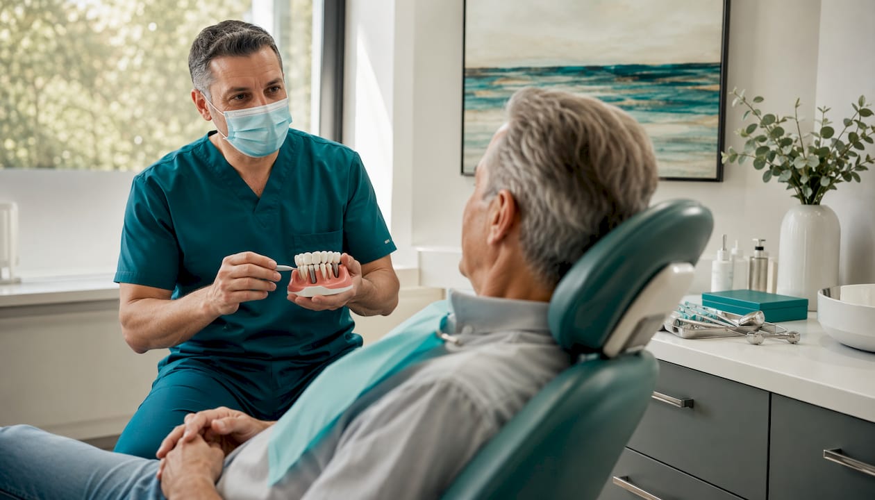
- What to expect at your consultation
- Key takeaways
- A clinical perspective on bone grafting
- Useful sources and further reading
- FAQ
How does a dental bone graft actually work?
Grafts act as a biological scaffold. Placed into a deficient area of the jaw, the material supports vascular ingrowth and triggers the body’s own bone-forming cells to populate the site, a process clinicians describe as osteoconduction and osteoinduction. Over weeks and months, the scaffold is gradually replaced by the patient’s own bone tissue.
The clinical indications are broader than many patients realise:
- Extraction socket preservation — placing graft material immediately after tooth removal to prevent the socket walls from collapsing inward.
- Ridge augmentation — rebuilding the height and width of a resorbed alveolar ridge to create a platform for implants.
- Sinus lift — elevating the maxillary sinus membrane and packing graft material beneath it when the upper posterior jaw lacks sufficient height.
- Trauma or infection-related bone loss — restoring volume lost to fracture, cyst removal, or advanced periodontal disease.
NHS guidance notes that bone grafting is a well-established procedure, and it has become a routine surgery worldwide in modern oral surgery.
What graft materials might your surgeon use?
The choice of material shapes the surgical plan, the recovery, and the biological outcome. There are five main categories:
- Autograft (your own bone): Harvested from the chin, jaw ramus, or, for larger defects, the hip or tibia. Biologically superior because it contains living cells and growth factors, but it requires a second surgical site. Hip harvests can limit walking for two to four weeks, a recovery burden patients often underestimate.
- Allograft (human donor bone): Processed cadaveric bone, carefully screened and sterilised. Avoids a donor-site operation and is widely used for moderate defects.
- Xenograft (animal-derived bone): Most commonly bovine. Its mineral composition closely resembles human bone, making it a reliable osteoconductive scaffold with a long clinical track record.
- Alloplast (synthetic materials): Calcium phosphate ceramics, hydroxyapatite, and bioactive glasses. Synthetic materials offer wide availability, no disease-transmission risk, and no donor site, though they generally lack the osteoinductive properties of autograft.
- Autologous tooth-derived biomaterials: An emerging category in which the patient’s own extracted tooth is processed into a graft scaffold. A 2025 systematic review reported 42–56% new bone formation at four to six months and implant survival rates of 96–100%, positioning this as a clinically credible alternative for suitable patients.
Growth factors such as BMP-2 are sometimes added to synthetic or xenogeneic scaffolds. Meta-analyses show modest gains in some width measures, but BMP-2 does not consistently increase new bone proportion or reduce complication rates, so its use warrants careful cost-benefit consideration.
Pro Tip: Ask your surgeon which material they recommend and why, whether a barrier membrane will be used, and whether any growth factors are included. Understanding the rationale helps you weigh the trade-offs before you consent.
What happens on the day of your procedure?
The exact steps depend on defect size, but the sequence follows a consistent clinical pattern:
- Pre-operative imaging: Cone beam CT (CBCT) confirms defect dimensions and guides graft volume planning.
- Anaesthesia: Local anaesthetic for minor in-clinic grafts; intravenous sedation or general anaesthetic for larger procedures performed in a hospital theatre.
- Tissue reflection: The surgeon makes an incision in the gum and lifts the soft tissue to expose the deficient bone.
- Graft placement: Material is packed or fixed into the defect. Block grafts from intraoral donor sites (symphysis or ramus) are secured with titanium screws; particulate grafts are held in place with a resorbable or non-resorbable membrane.
- Membrane and closure: A collagen or PTFE membrane is placed over the graft to exclude soft tissue and guide bone regeneration. The gum is sutured closed.
- Immediate post-operative checks: Bleeding control, bite assessment, and written aftercare instructions before discharge.
Adjuncts such as platelet-rich fibrin (PRF) or PRP are sometimes incorporated to support early healing. For complex reconstructions, full-arch dental implants may be planned as the final prosthetic stage once grafting is complete.
Healing, recovery, and warning signs to watch for
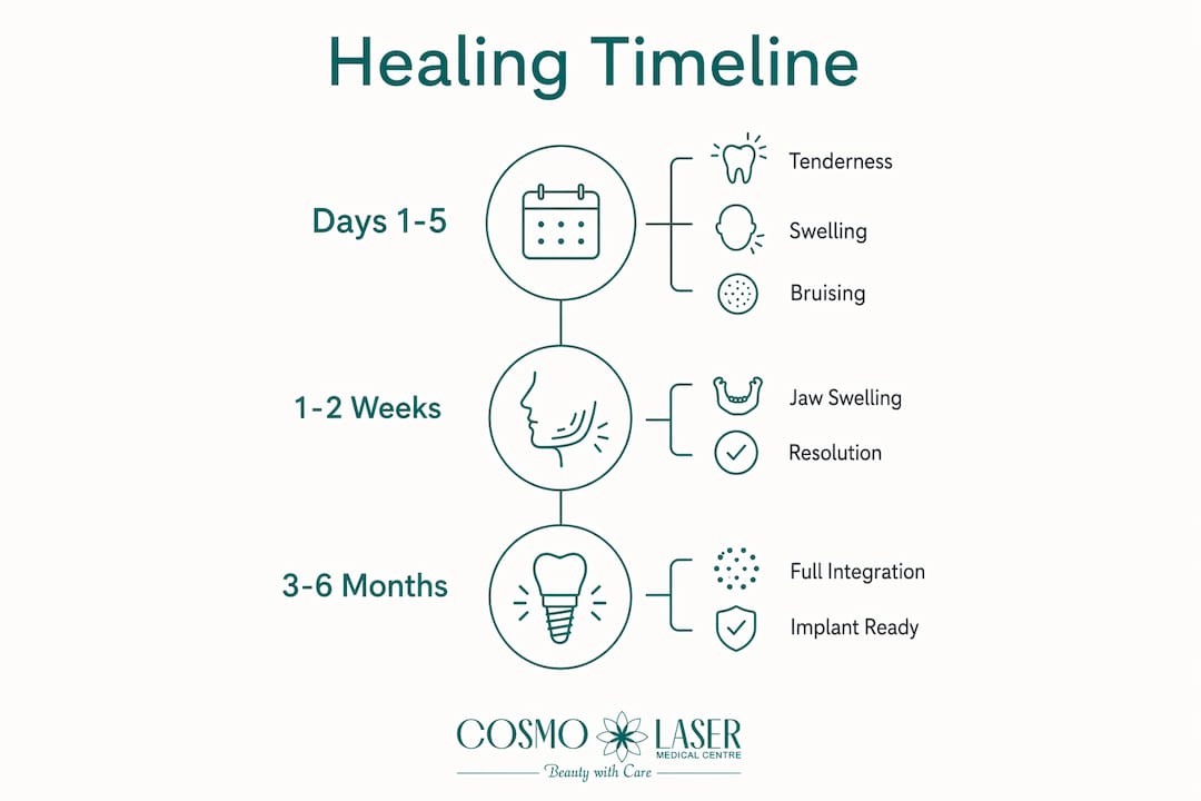
Initial recovery takes days to weeks; full graft integration is a longer process. Guy’s and St Thomas’ NHS Foundation Trust guidance places jaw swelling at 1–2 weeks and graft healing at 3–6 months before implant placement.
| Milestone | Typical timeframe |
|---|---|
| Tenderness, swelling, bruising | Days 1–5 |
| Jaw swelling resolves | 1–2 weeks |
| Suture removal (non-resorbable) | 10–12 days |
| Graft integration (minor grafts) | 3–6 months |
| Graft integration (major block grafts) | Up to 6 months or longer |
Some patients notice small, sand-like bone particles in the mouth in the first few days. This is normal in early recovery and not a sign of graft failure.
Aftercare essentials:
- Take prescribed analgesics (typically ibuprofen or paracetamol) on schedule for the first 48–72 hours rather than waiting for pain to peak.
- Complete any antibiotic course in full.
- Eat soft foods (yoghurt, soup, mashed vegetables) for at least two weeks; avoid hard, crunchy, or chewy foods near the graft site.
- Rinse gently with chlorhexidine mouthwash as directed; avoid vigorous rinsing or spitting for 24 hours post-surgery.
- No smoking. Nicotine constricts blood vessels and directly impairs graft healing.
- Avoid strenuous exercise for at least one week.
Contact your clinic promptly if you experience: increasing pain after day seven, fever above 38°C, persistent drainage or pus, or new numbness that does not resolve. Practical recovery guidance is also available from dental recovery specialists.
Risks, complications, and implant success after grafting
Grafting is generally safe and predictable, but patients deserve a clear-eyed view of the risks. NHS guidance confirms that implants placed into grafted bone carry a higher failure risk than those placed into natural bone, and infection prevention is therefore central to post-operative care.
| Risk or complication | Notes |
|---|---|
| Infection | Most common complication; managed with antibiotics and hygiene |
| Graft failure / resorption | May require repeat grafting before implant placement |
| Sensory disturbance | Temporary numbness near donor or recipient site |
| Membrane exposure | Can compromise regeneration; requires early clinical review |
| Implant failure in grafted bone | Higher than in native bone; smoking significantly increases risk |
Clinical evidence: A 2025 systematic review of autologous tooth-derived biomaterials reported implant survival rates of 96–100% at four to six months, with 42–56% new bone formation — results comparable to established graft materials.
Patient-related factors that raise failure risk include active smoking, uncontrolled diabetes, osteoporosis, and poor oral hygiene. If a graft fails, the site is debrided, allowed to heal, and re-grafted after approximately three months. Surgeons monitor progress through clinical examination and CBCT imaging, which confirms whether sufficient bone density has formed to support implant placement.
Are there alternatives to bone grafting?
Grafting is not always the only path. Several clinically validated strategies can reduce or eliminate the need for conventional augmentation:
Short implants (typically 6 mm or less) can be placed in resorbed ridges where conventional implants would require grafting, though they suit only moderate bone loss. Ridge splitting widens a narrow ridge by separating the cortical plates and placing graft material between them, avoiding a separate donor site. Guided bone regeneration (GBR) uses a membrane alone or with particulate graft to regenerate contained defects, and is well-suited to localised ridge deficiencies.
For patients with severe maxillary atrophy, zygomatic implants anchor into the cheekbone rather than the jaw, bypassing the need for sinus grafting entirely. They require specialist training and careful prosthetic planning, but can deliver fixed teeth without the extended healing of major grafts.
Pro Tip: For any case involving major extraoral grafting or zygomatic implants, a second opinion from an independent oral and maxillofacial surgeon is worth the time. Complex reconstructions benefit from more than one experienced perspective.
What affects the cost of a dental bone graft in the UK?
Prices vary considerably. The key cost drivers are:
- Graft material: Autograft procedures involving a separate donor-site operation cost more than allograft or synthetic options.
- Number of graft sites: Socket preservation at a single site is far less involved than bilateral sinus lifts combined with ridge augmentation.
- Setting: In-clinic procedures under local anaesthetic are significantly less expensive than hospital theatre cases under general anaesthetic.
- Imaging: CBCT scans are essential for planning but add to the overall cost.
- Membranes and growth factors: Titanium-reinforced membranes and PRF kits carry additional material costs.
- Follow-up visits and fixation screw removal: Some block graft protocols require a second minor procedure to remove titanium screws before implant placement.
Pro Tip: Request a fully itemised written estimate before committing. Confirm whether the quote includes consultations, imaging, theatre fees, all follow-up appointments, and any potential screw-removal procedure. Some clinics offer staged payment plans — ask about this at your first appointment.
How to choose the right clinic or surgeon
The surgeon’s experience with grafting specifically, not just implants in general, is the single most important factor. A clinician who places implants routinely but performs complex augmentation rarely is not the same as one who specialises in both.
What to check:
- GDC registration and any specialist listing in oral surgery or oral and maxillofacial surgery
- Volume of grafting procedures performed and willingness to share outcome data
- In-house CBCT imaging capability for accurate pre-operative planning
- A clear, written consent process that covers risks, alternatives, and contingency plans
- Defined post-operative support, including out-of-hours contact for urgent concerns
Questions worth asking at consultation:
- “Which graft material do you recommend for my case, and why?”
- “What are your complication and re-graft rates?”
- “Will I need a hospital stay, and which facility would you use?”
- “What happens if the graft does not integrate?”
Red flags: high-pressure sales tactics, no imaging before treatment planning, vague or verbal-only consent, and no clear plan for managing complications.
What to expect at your consultation
A thorough consultation covers clinical examination, CBCT imaging, a review of your medical history, and a stepwise treatment plan with realistic timelines. Arrive prepared:
- Bring a full list of current medications, including supplements and anticoagulants.
- Bring any existing dental X-rays or scans.
- Note your medical history clearly: diabetes, osteoporosis, bisphosphonate use, and cardiovascular conditions all affect surgical planning.
- Be honest about smoking status. Surgeons need accurate information to assess risk and advise on cessation support.
- Write down your goals and any concerns in advance so the consultation covers everything that matters to you.
A high-quality treatment plan will include a stepwise timeline from grafting through to final prosthetic placement, itemised costs, a contingency plan if the graft requires revision, and a scheduled follow-up programme. If you are considering implants alongside grafting, the dental implants guide at Cosmolaser Medical Centre provides further context on timelines and maintenance.
Key takeaways
Dental bone grafting makes implant treatment possible for many patients who would otherwise be unsuitable, but it demands careful planning, an experienced surgical team, and realistic expectations about healing time.
| Point | Details |
|---|---|
| Healing takes months, not weeks | Graft integration typically requires 3–6 months before implant placement can proceed. |
| Material choice matters | Autograft remains the gold standard; tooth-derived grafts show 96–100% implant survival in recent evidence. |
| Risks rise with lifestyle factors | Smoking and uncontrolled diabetes significantly increase graft failure risk. |
| Major grafts may need hospital admission | General anaesthesia and a short hospital stay are required for large-volume augmentation. |
| Get an itemised quote | Confirm that imaging, theatre fees, follow-ups, and screw removal are all included in the estimate. |
A clinical perspective on bone grafting
The conversation around dental bone grafting has shifted considerably in recent years, and not always in the direction patients expect. The arrival of tooth-derived biomaterials and refined GBR protocols has genuinely expanded what is achievable without a hip harvest, and that matters enormously for patient comfort and recovery planning. Yet the clinical fundamentals have not changed: a graft is only as good as the biological environment it is placed into.
What concerns me most in practice is the gap between what patients are told and what they genuinely understand before consenting. The 3–6 month healing window is not a formality. Placing an implant prematurely into incompletely integrated bone is one of the most avoidable causes of implant failure. Equally, delaying implant placement indefinitely after a graft has healed allows the new bone to shrink and lose density, creating a second problem where there was none. Timing is a clinical judgement, not a preference.
Patients considering complex augmentation deserve a surgeon who will show them their CBCT scan, explain the material rationale, and give them a written contingency plan. At Cosmolaser Medical Centre, the approach to dental planning is built on exactly that: personalised assessment, multidisciplinary input, and transparent communication at every stage. For complex cases or where major extraoral grafting has been proposed, a second opinion is not a sign of distrust. It is sound clinical practice.
To discuss your suitability for bone grafting or dental implants, book a consultation at Cosmolaser Medical Centre and request a CBCT assessment as part of your initial appointment.
Useful sources and further reading
- Bone grafting for dental implants, Guy’s and St Thomas’ NHS Foundation Trust — UK hospital trust guidance on procedure, anaesthesia, and recovery; the primary reference for NHS practice standards in this article.
- Autologous tooth-derived biomaterials in alveolar bone regeneration: a systematic review (MDPI, 2025) — the key clinical evidence on tooth-derived grafts, including the 42–56% new bone formation and 96–100% implant survival figures.
- Bone grafts in dental medicine: an overview of autografts, allografts and synthetic materials, PMC — peer-reviewed overview of all major graft material categories and their biological properties.
- Dental bone graft: process, purpose and healing, Cleveland Clinic — accessible patient-facing overview of the procedure and recovery.
- BMP-2 in oral-maxillofacial bone augmentation: a systematic review and meta-analysis — evidence on growth-factor-enhanced grafting and its limitations.
- CBCT imaging at Cosmolaser Medical Centre — cone beam CT planning service referenced in the consultation and imaging sections.
- Dental implants at Cosmolaser Medical Centre — clinic service page for patients progressing from graft planning to implant treatment.
This article provides general information for educational purposes only and does not constitute medical or dental advice. Confirm current clinical guidelines and your individual suitability with a qualified dental surgeon or oral and maxillofacial specialist.
FAQ
What is a dental bone graft used for?
A dental bone graft rebuilds lost or insufficient jawbone to allow dental implant placement or to restore jaw structure after tooth loss, trauma, or advanced periodontal disease.
How long does a dental bone graft take to heal?
Jaw swelling typically resolves within 1–2 weeks, but full graft integration usually takes 3–6 months before implant placement can proceed, according to Guy’s and St Thomas’ NHS Foundation Trust guidance.
Is a bone graft painful?
The procedure itself is performed under local anaesthetic or sedation, so patients feel pressure rather than pain. Post-operative discomfort is managed with standard analgesics and usually settles within the first week.
Can I get dental implants if I have bone loss?
Yes, in most cases. Bone grafting restores the volume needed to support an implant, though the extent of augmentation required depends on the degree of loss. Options such as implants with bone loss are discussed in detail by specialist dental teams.
Does smoking affect bone graft success?
Smoking significantly increases the risk of graft failure and implant failure in grafted bone. NHS guidance and clinical evidence both identify smoking as the most important modifiable risk factor, and cessation before surgery is strongly advised.