
TL;DR:
- Dark under-eye circles result from multiple factors such as pigment buildup, vascular translucency, structural hollows, or fluid retention. Proper treatment depends on identifying the underlying mechanism, with options ranging from topical agents to laser procedures or fillers. Prompt clinical review is necessary if the darkening affects one eye or worsens rapidly.
Black under-eye circles form when skin pigment, visible blood vessels, structural hollowing, or fluid accumulation — individually or in combination — create darkness in the infraorbital area. The PMC review on infraorbital dark circles confirms this is a multifactorial condition requiring an individualised approach, not a single-cause problem with a single fix. Most cases are not medically serious, but some warrant a GP or dermatology review.
Three immediate steps to take now:
- Apply a cold compress for five minutes to reduce vascular congestion and morning puffiness.
- Apply a broad-spectrum SPF to the orbital rim daily to prevent UV-driven pigmentary worsening.
- Check whether you have uncontrolled allergies or hay fever — chronic eye rubbing is a significant and often overlooked trigger.
When to see a clinician promptly: if the darkening affects only one eye, has appeared or worsened rapidly, or is accompanied by swelling, pain, or systemic symptoms, book a GP appointment rather than trying self-care first.
Table of Contents
- What are dark under-eye circles and how do the subtypes differ?
- The main black under-eye circles causes, grouped by mechanism
- Who is most likely to develop dark circles?
- How clinicians assess dark circles and when you need a review
- Practical home measures and cosmetic steps to try first
- Clinical and aesthetic treatment options for dark circles
- How to reduce recurrence and long-term risk
- Key research findings and clinical cautions
- Key takeaways
- A clinician’s perspective on realistic outcomes
- Useful sources for further reading
- FAQ
What are dark under-eye circles and how do the subtypes differ?
The term “dark circles” covers four distinct clinical mechanisms, and telling them apart matters because each responds to different treatments.
Pigmentary circles arise from excess melanin in the periorbital skin. They appear brown or grey-brown, do not change colour when you press the skin, and tend to be more prominent in brighter lighting. Sun exposure, post-inflammatory hyperpigmentation, and dermal melanocytosis are common drivers.
Vascular or translucency circles occur when thin lower-eyelid skin allows the underlying venous network and orbicularis oculi muscle to show through. The colour is typically blue-purple or pink-red. Pressing the skin lightly causes the colour to blanch partially, which distinguishes this type from pure pigment.

Structural or shadowing circles result from tear trough hollows, fat descent, and orbital rim recession. They look darker in overhead lighting and lighter in direct frontal light. No amount of topical cream corrects a structural hollow.
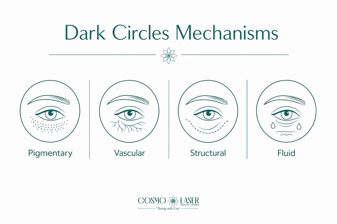
Fluid or oedematous circles involve periorbital fluid accumulation that creates a puffy lower lid casting a shadow. They are typically worse on waking and after salty meals. Multiple mechanisms often coexist, and appearance can fluctuate day to day depending on sleep, hydration, and posture.
The main black under-eye circles causes, grouped by mechanism
Understanding which mechanism is driving your dark circles is the most useful thing you can do before spending money on treatments. The PMC review groups causes into four biological pathways.
Pigmentary causes
- Dermal melanosis and melasma: excess melanin deposited in the dermis, often with a genetic or hormonal component.
- Post-inflammatory hyperpigmentation (PIH): repeated inflammation from atopy, contact dermatitis, or chronic eye rubbing triggers melanocyte activity, creating a self-sustaining darkening cycle that needs both trigger control and topical pigment treatment.
- UV exposure: cumulative sun damage accelerates periorbital pigmentation, particularly in Fitzpatrick skin types III–VI.
- Medication-related pigmentation: ophthalmic prostaglandin F2α analogues such as bimatoprost and latanoprost, used to treat glaucoma, are documented to increase periorbital pigmentation. Oral contraceptives can also contribute.
Vascular and translucency causes
The lower eyelid skin is among the thinnest on the body, with minimal subcutaneous tissue. This allows the underlying venous plexus and orbicularis oculi muscle to show through as a blue-purple tint. Haemoglobin breakdown products — specifically haemosiderin and biliverdin — can extravasate into periorbital tissues, creating a pigmentary depth that topical creams alone often cannot reverse.
Structural and shadowing causes
Age-related midface volume loss and ligamentous tethering create tear trough hollows that deepen shadows. As facial fat descends and orbital rim bone volume decreases, the relatively inflexible retaining ligaments tether the skin, creating a groove that overhead lighting accentuates. This is a structural problem, not a pigmentary one.
Fluid and oedema causes
Periorbital oedema is often worse after salty meals or on waking. Systemic fluid retention, atopy, and local venous or lymphatic insufficiency all contribute. The fluid takes on a purplish hue because of the prominent orbicularis muscle beneath.
Behavioural and lifestyle contributors
Sleep quality can affect the visibility of underlying vessels, potentially influencing the appearance of dark circles. Dehydration, smoking, and excess alcohol all impair skin quality and microcirculation. Allergic conjunctivitis drives chronic rubbing, which compounds both pigmentary and vascular mechanisms. Thyroid conditions and certain medications can also contribute.

Mechanism comparison table
| Mechanism | Typical appearance | First-line conservative measures |
|---|---|---|
| Pigmentary (melanin) | Brown/grey-brown; no blanching on pressure | SPF, topical vitamin C or niacinamide, allergy control |
| Vascular/translucency | Blue-purple; partial blanching on pressure | Cold compress, adequate sleep, head elevation |
| Structural/shadowing | Worse in overhead light; no colour change | Concealer; filler or fat transfer for correction |
| Fluid/oedema | Puffy, worse on waking; purplish tint | Head elevation, salt reduction, allergy treatment |
| Haemoglobin breakdown | Deep brown-purple; persistent | Specialist laser or peel; topical agents limited |
Who is most likely to develop dark circles?
Individuals with darker skin tones may experience periorbital hyperpigmentation more frequently than those with lighter skin tones. The PMC review and DermNet both note that pigmentary dark circles are particularly prevalent in people with Fitzpatrick skin types IV–VI, partly because melanocytes in these skin types respond more vigorously to inflammation and UV stimulation.
Clear risk factors include:
- Family history: a strong genetic component governs orbital anatomy, skin thickness, and melanocyte activity.
- Ageing: skin thinning, fat descent, and orbital rim recession worsen both vascular translucency and structural shadowing over time.
- Atopy and chronic rubbing: eczema and allergic rhinitis drive repeated periorbital inflammation, feeding the PIH cycle.
- Certain medications: glaucoma drops (prostaglandin analogues), oral contraceptives.
- Smoking: impairs collagen synthesis and microvascular health, accelerating skin thinning.
- Poor sleep and fluid retention: amplify vascular visibility and puffiness.
Age-related structural changes deserve particular mention; understanding how ageing affects periorbital tissues is explained in detail in the anti-wrinkle formulation research. Even in people with no pigmentary tendency, the tear trough hollow that develops from midface volume loss in the 30s and 40s can create the appearance of dark circles purely through shadowing.
How clinicians assess dark circles and when you need a review
A GP or dermatologist will typically begin with a detailed history: when the darkening started, whether it affects one or both eyes, whether it fluctuates, and what medications you take. The physical examination includes a press test (blanching indicates a vascular component), assessment under different lighting angles, and sometimes dermoscopy to characterise dermal versus epidermal pigment.
Blood tests are not routine but may be ordered if a systemic cause is suspected, such as thyroid dysfunction or iron deficiency.
Red flags that warrant prompt clinical review:
- Darkening affecting only one eye (unilateral change).
- Rapid or progressive worsening over weeks.
- Associated lump, skin ulceration, or persistent pain.
- Systemic symptoms such as fatigue, weight change, or eye pain.
Dark circles are rarely a sign of serious disease, but unilateral or rapidly progressive change should not be attributed to lifestyle without clinical assessment. At a specialist consultation, you may be referred to dermatology, ophthalmology or oculoplastics, or allergy services depending on the suspected cause. Knowing when to consult a dermatologist rather than managing alone can save considerable time and money.
Practical home measures and cosmetic steps to try first
For most people, a structured self-care approach is the right starting point before considering clinical procedures.
Short-term measures:
- Cold compress (wrapped ice or chilled spoons) for five minutes to constrict periorbital vessels and reduce puffiness.
- Sleep with your head slightly elevated to reduce overnight fluid pooling.
- Antihistamines or intranasal corticosteroids if allergic rhinitis is a confirmed trigger — treating the allergy reduces rubbing and inflammation.
- Consistent seven to nine hours of sleep; even one night of poor sleep visibly increases vascular translucency.
Daily skincare:
- A gentle, fragrance-free moisturiser to support the skin barrier and reduce inflammation risk.
- Dedicated SPF on the orbital rim every morning. Standard facial sunscreens are often avoided near the eyes; look for mineral formulations labelled as eye-safe.
- Topical vitamin C (L-ascorbic acid) or niacinamide can reduce melanin synthesis over several weeks of consistent use. Retinoids, used carefully at low concentrations, support collagen and can thin the appearance of vascular circles over time.
Caution with unregulated products: avoid unlicensed skin-lightening creams containing mercury or high-concentration hydroquinone, which are illegal in the UK and carry serious risks including exogenous ochronosis and paradoxical PIH, particularly in darker skin tones.
Pro Tip: For vascular or pigmentary circles, apply a peach or salmon-toned colour corrector before your concealer. The complementary tone neutralises blue-purple or brown discolouration far more effectively than concealer alone, and you need less product overall.
Clinical and aesthetic treatment options for dark circles
When self-care is insufficient, the treatment chosen should match the mechanism. A clinical review confirms that treatment must be individualised; no single modality addresses all subtypes.
Topical prescription agents: hydroquinone (available on prescription in the UK), azelaic acid, and tretinoin are first-line for pigmentary circles. They work over months, not weeks, and require sun protection to prevent rebound pigmentation.
Chemical peels: superficial to medium-depth peels using glycolic or trichloroacetic acid can reduce epidermal pigmentation. Chemical peels carry a PIH risk in darker skin tones and should be performed by a clinician experienced with higher Fitzpatrick types.
Laser and light devices: picosecond lasers and Q-switched Nd:YAG lasers target dermal melanin with lower thermal injury than older devices, reducing but not eliminating PIH risk. Vascular lasers (pulsed dye, KTP) address the vascular component. Laser treatments for periorbital pigmentation require a practitioner with specific experience in this anatomically delicate area.
Injectables: hyaluronic acid fillers placed in the tear trough correct structural hollowing and the associated shadow. Structural tear trough hollows respond predictably to volume augmentation, but require practitioner expertise to avoid irregularities. Platelet-rich plasma (PRP) injections stimulate collagen and may improve skin quality and vascular appearance over a course of sessions.
Surgical options: blepharoplasty and fat repositioning address significant fat prolapse and structural laxity. These are considered when non-surgical options have been exhausted or when the structural component is pronounced.
UK clinical pathway: most patients begin with a GP consultation. The NHS will investigate and treat an underlying medical cause (e.g. thyroid disease, atopy) but does not fund elective cosmetic procedures for dark circles. Aesthetic treatments are accessed privately. Readers considering procedural options can review the cosmetic dermatology procedures checklist to understand what to expect before a consultation.
| Mechanism | Most effective clinical options |
|---|---|
| Epidermal/dermal melanin | Topical agents, chemical peels, picosecond or Q-switched laser |
| Vascular/translucency | Vascular laser, PRP, skin-quality improvement |
| Structural hollowing | Hyaluronic acid filler, fat transfer, blepharoplasty |
| Haemoglobin breakdown products | Specialist laser; topical agents have limited effect |
How to reduce recurrence and long-term risk
Prevention is most effective for pigmentary and fluid-related causes. Structural changes from ageing are not preventable with creams, but their rate of progression can be slowed.
Prevention checklist:
- Apply a mineral SPF to the orbital rim every morning, even in winter.
- Wear broad-brimmed sunglasses outdoors to shield the periorbital skin from UV scatter.
- Treat allergic conjunctivitis or eczema promptly to break the rubbing-PIH cycle.
- Moderate salt intake, particularly in the evening, to reduce morning puffiness.
- Avoid smoking; it accelerates collagen breakdown and skin thinning around the eyes.
- Maintain consistent sleep habits and stay well hydrated.
Pro Tip: Sunglasses are one of the most underused periorbital protection tools. UV scatter from pavements and water reaches the lower eyelid even when you are not looking directly at the sun. A wraparound or large-lens style provides meaningfully better coverage than standard frames.
Structural causes such as tear trough hollowing and orbital rim recession are driven by bone and fat volume loss that lifestyle measures cannot reverse. For these, volume augmentation remains the most predictable option when the time comes.
Key research findings and clinical cautions
The central conclusion of the PMC infraorbital dark circles review is that this condition is multifactorial and that a customised management strategy, built around the specific anatomic changes present, produces the best outcomes. No single treatment works for all subtypes.
Two clinical cautions deserve emphasis. First, people with darker skin tones face a higher risk of PIH from lasers and chemical peels, which means the risk-benefit calculation for these procedures differs significantly from that in lighter skin types. Specialist experience with higher Fitzpatrick types is not optional; it is a patient safety requirement. Second, where haemoglobin breakdown products such as haemosiderin and biliverdin have accumulated in the periorbital tissues, topical creams have limited reach. Specialist laser or procedural intervention is needed, and expectations should be set accordingly.
Medication-related pigmentation from ophthalmic prostaglandin analogues is a reversible cause that is frequently missed. If you use glaucoma eye drops and have noticed progressive periorbital darkening, discuss this with your prescriber before pursuing cosmetic treatment.
Key takeaways
Under-eye darkness is almost always multifactorial, and matching the treatment to the mechanism is the single most important step toward a useful outcome.
| Point | Details |
|---|---|
| Four distinct mechanisms | Pigmentary, vascular/translucency, structural shadowing, and fluid each require different treatment approaches. |
| Darker skin tones carry higher PIH risk | Lasers and peels must be selected and performed by a clinician experienced with higher Fitzpatrick skin types. |
| Structural causes need procedures | Tear trough hollowing from volume loss does not respond to topical creams; filler or surgery is required. |
| Red flags need prompt review | Unilateral, rapidly progressive, or symptom-associated darkening warrants GP assessment, not self-care. |
| Medication is a reversible cause | Glaucoma drops (prostaglandin analogues) can cause periorbital pigmentation; discuss with your prescriber. |
A clinician’s perspective on realistic outcomes
The most common mistake people make with dark circles is choosing a treatment before identifying the mechanism. A patient with structural tear trough hollowing who spends months on vitamin C serums will see little change, not because the product is poor, but because it cannot fill a volume deficit. Equally, someone with vascular translucency who pursues pigment lasers is targeting the wrong tissue layer entirely.
At Cosmolaser Medical Centre, the assessment always begins with mechanism identification: press test, lighting assessment, and a detailed history that includes medications, allergy status, and sleep patterns. Only then is a treatment plan discussed. For complex or treatment-resistant cases, a multi-modal approach combining topical agents, energy-based devices, and volume correction often produces results that no single modality achieves alone. Safety, particularly PIH risk in darker skin tones, is a non-negotiable part of that planning. If you are uncertain whether your dark circles warrant professional assessment, the answer is almost always yes — a single consultation clarifies the mechanism and prevents months of misdirected self-treatment.
This article provides general information only and is not a substitute for professional medical advice. Consult a GP or qualified dermatologist for guidance specific to your situation.
Useful sources for further reading
The following authoritative resources provide reliable clinical detail on dark circles, their causes, and UK-relevant care pathways:
- Infraorbital Dark Circles: A Review of the Pathogenesis, Evaluation and Treatment (PMC) — the most comprehensive peer-reviewed review of causes and treatment modalities.
- Dark circles under the eyes — DermNet — clinically accurate overview with particular attention to pigmentary causes and skin tone considerations.
- Dark circles under eyes — Mayo Clinic — accessible summary of causes and when to seek clinical review.
- Bags under eyes: symptoms and causes — Mayo Clinic — explains the structural and fluid mechanisms behind lower-eyelid puffiness.
- Dark circles under the eyes: causes and treatments — Medical News Today — broad overview including lifestyle, medical, and cosmetic factors.
- Dark circles under eyes — Cleveland Clinic — clinical guidance on when to escalate to specialist review.
FAQ
What can dark circles under the eyes be a symptom of?
Dark circles are rarely a sign of serious disease. They most commonly reflect genetics, ageing, sleep patterns, allergies, or skin pigmentation rather than an underlying medical condition. Unilateral or rapidly progressive darkening, however, should be assessed by a GP to rule out local or systemic causes.
What deficiency causes dark circles under the eyes?
Iron deficiency can contribute to periorbital pallor and vascular visibility, and vitamin K deficiency has been linked to impaired haemoglobin clearance in periorbital tissues. That said, deficiency is one of many possible contributors, not the primary cause in most adults.
How do you get rid of black under-eye circles?
Treatment depends on the mechanism. Pigmentary circles respond to topical agents (vitamin C, niacinamide, retinoids) and specialist laser or chemical peels. Structural hollowing requires filler or surgery. Vascular circles improve with cold compresses, sleep, and vascular laser. No single product or procedure works for all types.
Do dark circles under the eyes mean liver problems?
No. Dark circles are not a recognised marker of liver disease. While severe liver conditions can cause skin discolouration elsewhere on the body, periorbital darkening is overwhelmingly caused by pigment, vascular translucency, structural anatomy, or lifestyle factors rather than hepatic dysfunction.