
What is CBCT and why is it used in clinical practice?
Cone beam computed tomography, universally known as CBCT, is a specialised 3D X-ray imaging technology that captures volumetric data of teeth, bone, and nerve pathways in a single rotation of the scanner around the patient’s head. Unlike conventional 2D dental X-rays, which compress three-dimensional anatomy into a flat image, CBCT produces sub-millimetre resolution images that clinicians can view in axial, coronal, and sagittal planes. The result is a level of anatomical clarity that fundamentally changes what is possible in diagnosis and treatment planning.
Traditional panoramic or periapical X-rays remain useful for routine assessments, but they carry inherent limitations: magnification, distortion, and structural superimposition. CBCT eliminates all three. Where a 2D image might obscure the precise position of a nerve canal or the true extent of a cyst, a CBCT scan renders each structure in its exact spatial relationship to surrounding anatomy.
Key reasons clinicians indicate CBCT:
- Precise localisation of nerve pathways before implant or surgical procedures
- Assessment of bone volume, density profile, and alveolar architecture
- Detection of root canal morphology that conventional radiographs miss
- Evaluation of impacted or supernumerary teeth and their proximity to vital structures
- Diagnosis of temporomandibular joint (TMJ) disorders and jaw pathology
- Identification of cysts, tumours, and pathological calcifications
- Orthodontic skeletal analysis in complex treatment cases
Table of Contents
- What happens during a CBCT scan and what should you expect?
- How CBCT is used across dentistry and aesthetic medicine
- Radiation exposure and safety guidelines in the UK
- Why choose a clinic with CBCT capability for your treatment?
- What happens after your CBCT scan?
- What does a CBCT scan cost in the UK?
- How are CBCT images interpreted and analysed?
- Limitations and contraindications of CBCT
- Key takeaways
- FAQ
What happens during a CBCT scan and what should you expect?
The procedure is straightforward and entirely non-invasive. No special preparation is required beyond removing metal objects such as jewellery, glasses, and removable dental appliances before the scan begins. You remain fully clothed throughout.
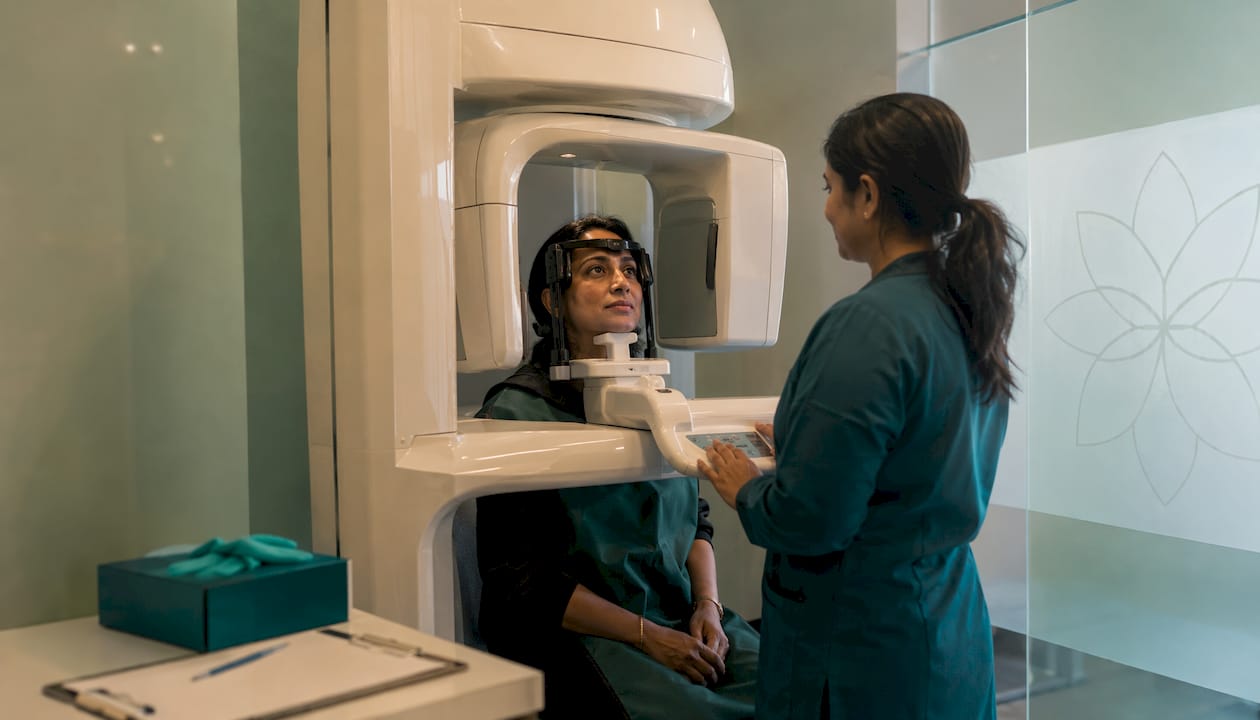
You will be positioned standing or seated within the CBCT unit, with your head stabilised to minimise movement. The scanner arm rotates around your head, capturing multiple two-dimensional projection images within a single sweep. For a full-mouth scan, the acquisition typically takes seconds; targeted small-field scans complete in considerably less time. The captured images are reconstructed automatically into a three-dimensional digital volume, available for clinical review within minutes.
In-clinic CBCT machines mean you avoid referral delays to external radiology centres, and your clinician can review findings with you on the same visit.
Patient expectations at a glance:
- No injections, no sedation, and no physical discomfort
- Metal objects removed prior to positioning
- Scan duration of 20–40 seconds for full-arch imaging
- Immediate 3D reconstruction available for same-appointment review
- Results interpreted by a trained clinician before any treatment decision is made
How CBCT is used across dentistry and aesthetic medicine
CBCT is the gold standard for implant dentistry, endodontic assessment, and the localisation of impacted teeth, owing to the precision of its three-dimensional data. For dental implant planning, it reveals the exact height and width of available bone, the proximity of the inferior alveolar nerve, and the density profile of the alveolus — information that directly reduces the risk of implant failure or nerve injury.
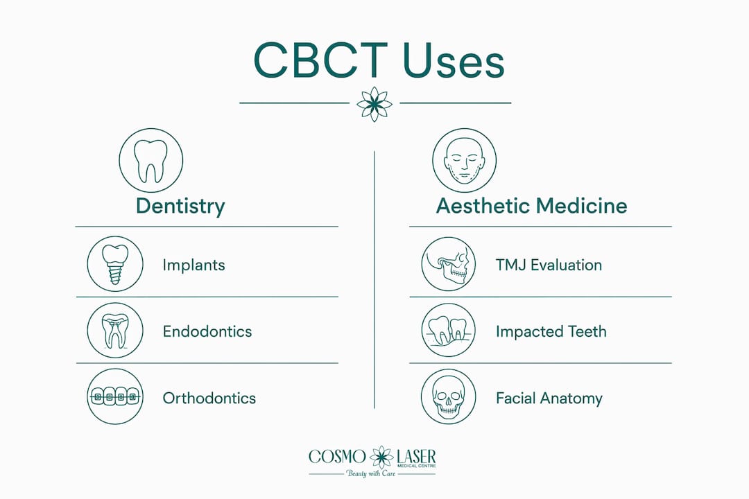
In endodontics, CBCT reveals root canal morphology that panoramic or periapical films routinely miss: accessory canals, calcified chambers, root fractures, and periapical pathology. Clinicians planning root canal treatment rely on this detail to avoid procedural errors. Orthodontic applications include skeletal and dental structure analysis for patients with impactions, asymmetries, or complex malocclusions where 2D cephalometry is insufficient.
TMJ assessment benefits considerably from CBCT’s ability to image condylar morphology, joint space, and bony changes associated with degenerative disorders. Oral and maxillofacial surgeons use it to plan the removal of impacted third molars, assess midfacial fractures, and delineate the extent of odontogenic cysts or tumours. One important caveat: CBCT is less effective than medical CT at evaluating soft tissue pathologies, where conventional CT or MRI remains the appropriate modality.
Primary clinical scenarios where CBCT improves outcomes:
- Pre-surgical implant site assessment and guided surgery planning
- Complex endodontic diagnosis and retreatment evaluation
- Orthodontic impaction localisation and skeletal analysis
- TMJ condylar assessment and degenerative joint disease staging
- Surgical planning for impacted canines and third molars
- Jaw cyst, tumour, and pathological calcification delineation
- Post-operative bone graft and implant position verification
Radiation exposure and safety guidelines in the UK
CBCT involves more radiation than conventional 2D dental X-rays, though total radiation doses from CBCT are 96% lower than those from conventional medical CT examinations. UK clinical practice is governed by the principle that every scan must be justified: diagnostic benefit must demonstrably outweigh radiation risk before a scan is authorised.
Two guiding principles frame this: ALARA (as low as reasonably achievable) and ALADA (as low as diagnostically acceptable). UK guidelines require clinicians to select the smallest appropriate field of view (FOV) for each clinical task. A small FOV targeted at a single tooth delivers a fraction of the dose produced by a large FOV covering the entire maxillofacial region. Small FOV protocols are particularly well-suited to root canal morphology assessment and impacted tooth localisation, avoiding unnecessary irradiation of surrounding structures.
Radiation safety measures applied in UK practice:
- Justification of every scan against clinical need before exposure
- Selection of the smallest FOV adequate for the diagnostic task
- Use of low-dose and ultra-low-dose acquisition protocols
- Appropriate mA and kVp settings to minimise absorbed dose
- Patient thyroid and gonad shielding where applicable
- Restriction of CBCT to cases where 2D imaging is insufficient
Why choose a clinic with CBCT capability for your treatment?
Access to in-house CBCT transforms the quality and speed of clinical decision-making. At Cosmolaser Medical Centre, CBCT imaging is integrated directly into the diagnostic and treatment planning workflow, meaning patients receive precise, same-visit assessment without referral delays. The qualified clinical team adheres to UK guidelines on scan justification, FOV selection, and dose optimisation, ensuring every scan is both clinically necessary and performed at the lowest appropriate radiation level.
Expert consensus confirms that trained clinicians who understand imaging protocols and interpretation accuracy are central to safe CBCT use. Cosmolaser Medical Centre’s approach to combination aesthetic and dental treatment is built on precisely this foundation: advanced technology, qualified specialists, and a patient-centred protocol that tailors every scan to individual anatomy and clinical need.
Clinic credentials and patient benefits:
- In-house CBCT unit eliminating referral delays
- Qualified clinicians trained in CBCT protocol optimisation and image interpretation
- Adherence to ALARA and ALADA principles on every scan
- Low-dose protocols selected according to clinical task
- Integrated dental and aesthetic treatment planning under one roof

What happens after your CBCT scan?
Once the scan is complete, the three-dimensional dataset is reviewed by the treating clinician, who assesses spatial relationships, bone quality, canal morphology, and any pathological signs in the context of your clinical presentation. CBCT interpretation requires integrating radiographic findings with your symptoms, clinical examination, and treatment objectives — it is never read in isolation.
Your clinician will discuss findings with you, explain what the images show, and outline the recommended treatment pathway. If the scan reveals findings outside the dental or maxillofacial scope, referral to an appropriate specialist is arranged. Follow-up imaging, where indicated, is scheduled at the minimum interval clinically justified.
What does a CBCT scan cost in the UK?
CBCT scans are more affordable and accessible than medical CT, though costs vary by clinic, FOV size, and clinical indication. Prices across UK private dental and medical clinics typically reflect the complexity of the scan and whether specialist reporting is included. Clinics with in-house equipment generally offer more competitive pricing than those relying on external radiology referrals, and the absence of a separate appointment reduces both time and indirect cost for patients.
NHS provision of CBCT is limited and largely restricted to secondary care settings such as oral and maxillofacial surgery departments. Most patients accessing CBCT for implant planning, endodontic assessment, or orthodontic evaluation do so through private clinics.
How are CBCT images interpreted and analysed?
CBCT datasets are viewed using dedicated software that allows the clinician to scroll through axial, coronal, and sagittal slices, generate cross-sectional views of specific anatomical regions, and produce three-dimensional surface renderings. For implant planning, cross-sectional views at the proposed implant site reveal bone height, width, and the precise position of the inferior alveolar canal. For endodontic cases, high-resolution small-FOV images display root canal anatomy at sub-millimetre detail.
Accurate interpretation demands both technical training and clinical experience. Findings are always contextualised against the patient’s presenting complaint and clinical examination, not reported as standalone radiographic observations.
Limitations and contraindications of CBCT
CBCT is not appropriate for every clinical situation. Its primary limitation is soft tissue imaging: it provides limited contrast resolution for muscles, nerves, and vascular structures, where conventional CT or MRI is superior. Movement artefacts can degrade image quality, particularly in patients who cannot remain still during the scan. Bone density values from CBCT cannot be directly correlated with Hounsfield units derived from medical CT, which limits their use in certain systemic assessments.
Relative contraindications include pregnancy, where ionising radiation is avoided unless clinically urgent, and patients with certain implanted electronic devices where positioning within the unit may be problematic. Children and adolescents require particular justification given their greater radiosensitivity, and small-FOV, low-dose protocols are prioritised in younger patients.
Key takeaways
CBCT delivers three-dimensional anatomical detail that 2D dental X-rays cannot provide, making it the preferred imaging modality for implant planning, endodontics, and complex surgical cases in UK clinical practice.
| Point | Details |
|---|---|
| 3D imaging advantage | CBCT captures volumetric data of teeth, bone, and nerves that conventional 2D X-rays cannot resolve. |
| Radiation context | CBCT doses are 96% lower than conventional medical CT, though higher than standard 2D dental X-rays. |
| Scan duration | Full-mouth CBCT acquisition typically takes 20–40 seconds; three-dimensional reconstruction is ready for review within minutes. |
| Safety principles | UK practice requires ALARA and ALADA compliance, with FOV selected to match the specific clinical task. |
| Soft tissue limitation | CBCT is less effective than medical CT or MRI for soft tissue evaluation; alternative imaging may be needed. |
FAQ
What is CBCT used for in dentistry?
CBCT is used for implant planning, endodontic assessment, orthodontic analysis, TMJ evaluation, and the localisation of impacted teeth, providing three-dimensional detail that 2D X-rays cannot deliver.
Is a CBCT scan safe?
Yes. CBCT doses are 96% lower than conventional medical CT, and UK clinical guidelines require every scan to be justified by diagnostic need, with the smallest appropriate field of view selected to minimise exposure.
Do I need to prepare before a CBCT scan?
No special preparation is required. You will be asked to remove metal objects such as jewellery, glasses, and removable dental appliances before the scan begins.
How long does a CBCT scan take?
The image acquisition for a full-mouth scan typically takes 20–40 seconds, and the three-dimensional reconstruction is ready for clinical review within minutes.
Can CBCT image soft tissues?
CBCT is optimised for hard tissue imaging. It provides limited soft tissue contrast, and conventional CT or MRI is recommended when soft tissue pathology is the primary diagnostic concern.