
Save your natural tooth, end the pain. CBCT-guided root canal treatment performed by experienced endodontic specialists at Cosmolaser Medical Centre — precise, gentle, and same-day where possible.
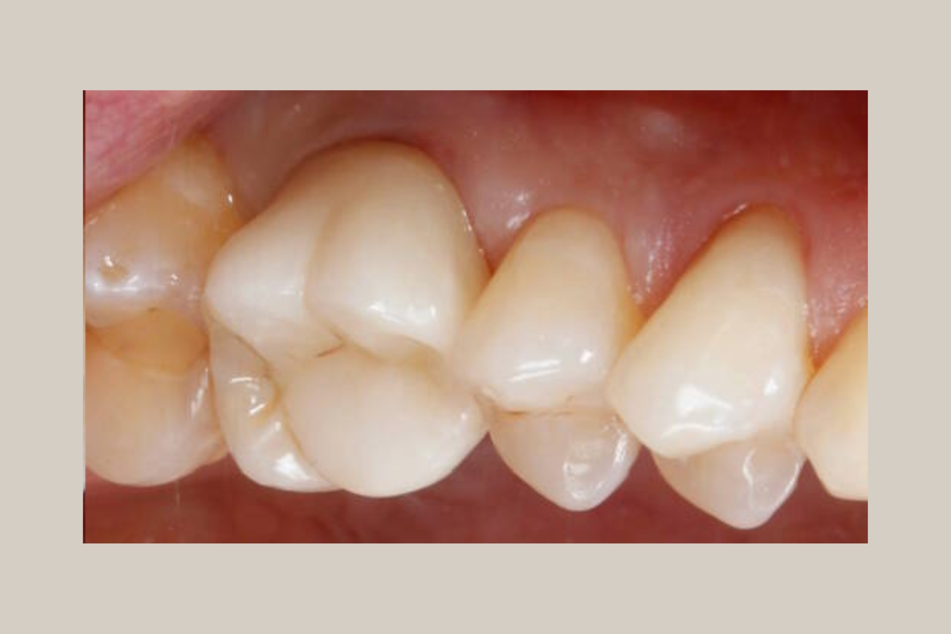
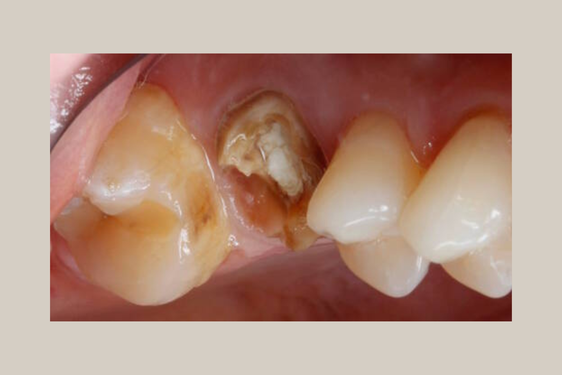
Root canal treatment has an undeserved reputation. For most patients treated with modern techniques and appropriate anaesthesia, a root canal is no more uncomfortable than having a filling placed — and it eliminates the severe, throbbing pain of a dental abscess or infected pulp that brought them to us in the first place. The procedure saves your natural tooth, which is always the preferred clinical outcome over extraction.
In Sharjah and across the UAE, dietary habits — including high sugar intake, frequent consumption of hot and cold beverages, and the teeth-grinding (bruxism) that many residents experience as a stress response — make pulp infection and tooth pain among the most common dental presentations we see. When a tooth’s nerve becomes infected, there is no home remedy that resolves it: the infection will worsen, spread to surrounding bone, and in serious cases require hospitalisation. Early root canal treatment is almost always simpler, faster and less costly than leaving an infection to progress.
At Cosmolaser Medical Centre, our dental team uses the in-house CBCT (Cone Beam CT) scanner to capture a full 3D image of your tooth’s root anatomy before treatment begins. Standard dental X-rays are 2D — they can miss accessory canals, curved root paths and periapical lesions that a 3D scan reveals clearly. This pre-treatment imaging allows us to plan the procedure with precision that simply is not possible with conventional radiography alone, reducing treatment time and the risk of complications.
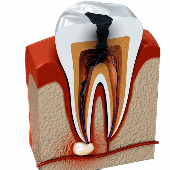
Root canal treatment (also called endodontic treatment) is a dental procedure that removes infected, inflamed or dead tissue from inside the tooth — specifically from the pulp chamber and root canals — cleans and disinfects the space, and seals it permanently to prevent re-infection. The tooth’s crown (the visible part) is preserved entirely; only the internal nerve tissue and blood supply are removed.
The pulp is the soft tissue at the centre of every tooth containing blood vessels, nerves and connective tissue. It is essential during tooth development, but a fully formed adult tooth can function perfectly without it — drawing nutrients from the surrounding periodontal ligament instead. Once the pulp becomes infected or necrotic, it cannot heal itself. Root canal treatment is the only way to save the tooth without extraction.
Before any root canal procedure at Cosmolaser, we use our in-house CBCT (Cone Beam Computed Tomography) scanner to capture a full 3D image of the affected tooth and surrounding bone. This is the most significant advancement in endodontic diagnosis of the past two decades.
A standard 2D periapical X-ray shows the tooth in a single plane. It can miss: additional root canals (upper molars commonly have 4 canals, but anatomy varies), curved or calcified canal paths that change the instrument approach, the true extent of periapical bone infection, and cracks or vertical root fractures invisible in 2D. Our CBCT scan reveals all of this before the first instrument is placed — allowing complete treatment planning, appropriate session allocation and a more predictable outcome.
Standard 2D X-ray (Periapical) | CBCT 3D Scan (Cosmolaser) |
|---|---|
Single-plane 2D image only | Full 3D volumetric image of tooth and bone |
May miss accessory canals | All root canals visible in all planes |
Limited periapical lesion assessment | Precise lesion size and extent mapped |
Cannot detect vertical root fractures | Fractures often detectable pre-treatment |
Canal curvature estimated | Exact canal curvature measured and planned for |
Available at most dental clinics | In-house at Cosmolaser — no referral needed |
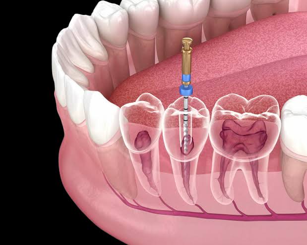
The standard root canal procedure involves accessing the pulp chamber through the crown of the tooth, removing all infected or necrotic tissue from the canals using a combination of rotary nickel-titanium instruments and irrigation with sodium hypochlorite and EDTA, shaping the canals to receive a permanent fill, and sealing them with gutta-percha and root canal sealer. A temporary or permanent crown is placed over the tooth afterwards to restore function and protect the treated structure.
Straightforward single-rooted teeth (incisors, canines, premolars) are often completable in a single session of 60–90 minutes. Multi-rooted teeth with complex anatomy (upper molars, teeth with calcified canals) or those with significant abscess requiring inter-appointment medication are managed across two sessions.
Sessions | 1 session (simple) or 2 sessions (complex / abscess) |
Duration Per Session | 60–120 minutes depending on tooth and complexity |
Anaesthesia | Local anaesthetic injection — highly effective; most patients feel no pain |
Pre-treatment Imaging | In-house CBCT 3D scan for all cases at Cosmolaser |
Instruments | Rotary nickel-titanium (NiTi) files — more efficient and less stressful on canals than manual stainless steel |
Canal Fill Material | Gutta-percha with biocompatible root canal sealer |
Post-treatment Restoration | Temporary filling at first visit; permanent crown recommended within 4–6 weeks |
A small percentage of root-canal-treated teeth fail over time — either due to missed canals at the original treatment, new decay breaking the seal, or persistent infection that did not fully resolve. Root canal retreatment involves reopening the tooth, removing the existing gutta-percha fill, thoroughly re-cleaning and reshaping all canals, and resealing the root system.
Retreatment is significantly more complex than a first-time procedure. CBCT imaging is particularly valuable here — it allows us to identify exactly why the original treatment failed, whether missed canals, persistent periapical pathology, or a vertical root fracture (in which case retreatment cannot save the tooth and extraction may be necessary). We assess retreatment feasibility honestly at consultation and will always present alternatives if retreatment is not the appropriate course.
Indication | Previously root-treated tooth with persistent pain, swelling or radiographic failure |
Sessions | Typically 2 sessions |
CBCT Imaging | Mandatory — required to assess cause of failure before retreatment |
Complexity | Higher than primary treatment — specialist assessment recommended |
Prognosis | Good when failure cause is identifiable and correctable; guarded for vertical fractures |
In cases where the pulp is partially inflamed but not fully infected — often seen in teeth with deep decay where the cavity approaches but has not fully penetrated the pulp — vital pulp therapy (pulpotomy or direct pulp capping) may allow the tooth’s nerve to be preserved without full root canal treatment. A biocompatible material (such as mineral trioxide aggregate, MTA) is placed directly over the exposed pulp tissue to encourage healing and maintain pulp vitality.
This is particularly relevant for younger patients whose root development may not be complete, and for patients where minimising intervention is the clinical priority. Success rates depend on the degree of pulp inflammation at the time of treatment — CBCT helps assess pulp chamber status accurately before this decision is made.
Indication | Partial pulp exposure with healthy remaining pulp tissue |
Sessions | Usually 1 session |
Material | MTA (Mineral Trioxide Aggregate) or Biodentine |
Advantage | Preserves tooth vitality — avoids full root canal treatment |
Follow-up | Radiographic review at 6 and 12 months to confirm healing |
The appropriate treatment is determined by the extent of pulp involvement, tooth anatomy, restorability of the crown, and patient factors. The CBCT scan and clinical examination at consultation provide the full picture needed to make this decision:
Clinical Situation | Recommended Approach | Notes |
|---|---|---|
Deep decay, pulp exposed, healthy remaining pulp | Vital pulp therapy (pulpotomy/capping) | Assessed by CBCT — preserves tooth vitality if successful |
Severe toothache, hot/cold sensitivity persisting | Full root canal treatment — 1 or 2 sessions | Most common presentation; high success rate |
Abscess / swelling / gum boil present | Root canal with inter-appointment medication | Antibiotic prescription often concurrent; 2 sessions standard |
Dead (necrotic) tooth, no acute pain | Root canal treatment | Often single session; no anaesthesia challenge as nerve non-vital |
Previously treated tooth failing | Root canal retreatment | CBCT mandatory; complexity and prognosis assessed first |
Vertical root fracture confirmed on CBCT | Extraction discussed | Retreatment cannot save fractured roots — honest assessment given |
Facial swelling / fever / difficulty swallowing | Emergency dental care — same day | Dental emergency; WhatsApp us immediately |
Root canal treatment can be performed on any tooth in the mouth. Complexity and session count vary by tooth anatomy:
Tooth Type | Typical Canal Count | Complexity | Notes |
|---|---|---|---|
Upper / Lower Incisors | 1 canal | Low | Straightforward; often single-session |
Canines | 1 canal | Low | Longest root in the mouth; simple canal path |
Upper Premolars | 1–2 canals | Low–Moderate | 2 canals common; CBCT confirms anatomy |
Lower Premolars | 1–2 canals | Low–Moderate | Canal variation possible; CBCT guides approach |
Upper First Molars | 3–4 canals (MB2 often missed on 2D X-ray) | High | Most complex upper tooth — CBCT essential for MB2 detection |
Upper Second Molars | 3–4 canals | High | Similar to upper first molar |
Lower Molars | 2–4 canals (C-shaped canal variants in UAE population) | High | C-shaped canal anatomy more prevalent in South/East Asian patients |
Wisdom Teeth | Variable — 1–5 canals | Very High | Root canal rarely recommended; extraction often preferred |
Each case below documents a representative clinical scenario treated at Cosmolaser Medical Centre. Radiographic images (CBCT axial/sagittal slices + periapical X-rays at pre-treatment, post-obturation and 6-month review) should accompany each case.
Male patient, 38 years. Severe spontaneous toothache, sensitivity to cold persisting >30 seconds. No swelling.
| Findings | ||
|---|---|---|
|
CBCT: 4-canal anatomy confirmed (MB1, MB2, DB, P). Periapical: deep mesial decay approaching pulp. No periapical pathology. |
Treatment: 1-session root canal. All 4 canals located and instrumented (NiTi rotary). Full gutta-percha obturation. Temporary crown placed. |
Outcome: Pain-free within 24 hours. Crown fitted at 4 weeks. 6-month CBCT: periapical bone normal. |
Female patient, 44 years. Swelling on lower left jaw, gum boil present. Tooth non-responsive to cold testing (necrotic pulp). Previous attempted filling 6 months prior.
| Findings | ||
|---|---|---|
|
CBCT: Periapical abscess confirmed on mesial root. 4-canal anatomy. Existing restoration incomplete — caries remaining. |
Treatment: 2-session root canal with calcium hydroxide inter-appointment dressing. Antibiotic prescribed (amoxicillin). Gutta-percha obturation at session 2. Crown referral. |
Outcome: Swelling resolved by session 2. Abscess healed radiographically at 3-month review. Crown placed, tooth fully functional. |
Female patient, 31 years. Discomfort and gum pimple on previously root-treated upper front tooth (treated 4 years prior elsewhere).
| Findings | ||
|---|---|---|
|
CBCT: Periapical lesion 4mm diameter. Existing gutta-percha fill short of apex by 3mm. Single canal — no missed canals. |
Treatment: Retreatment: existing fill removed, canal thoroughly cleaned and reshaped, MTA apical plug placed, gutta-percha obturation. Crown replaced. |
Outcome: Gum pimple resolved within 2 weeks. 12-month CBCT: periapical lesion fully resolved. Tooth asymptomatic. |
Male patient, 29 years (South Asian). Toothache and sensitivity to biting. Standard 2D X-ray at prior clinic inconclusive on canal anatomy.
| Findings | ||
|---|---|---|
|
CBCT: C-shaped canal morphology confirmed — single ribbon-shaped canal rather than discrete separate canals. Periapical lesion on distal root. |
Treatment: 1-session root canal using C-shape-specific irrigation protocol and lateral condensation obturation. Treatment planned entirely from CBCT data. |
Outcome: No complications. Standard protocol would have risked incomplete cleaning of the C-shaped isthmus. CBCT allowed complete debridement. Asymptomatic at 6 months. |
Female patient, 52 years. Chronic dull ache, previously root-treated lower molar. Retreatment requested.
| Findings | ||
|---|---|---|
|
CBCT: Vertical root fracture confirmed on mesial root — clearly visible in all three planes. Periapical lesion adjacent to fracture line. |
Treatment: Retreatment contraindicated. Extraction of fractured tooth discussed and performed. Implant placement planned after bone healing. |
Outcome: Informed decision enabled by CBCT diagnosis. Avoided unnecessary retreatment procedure. Implant placed 4 months post-extraction. |
Male patient, 22 years. Deep decay upper premolar. No spontaneous pain, mild sensitivity. Pulp partially exposed during decay removal.
| Findings | ||
|---|---|---|
|
CBCT: Root development complete. Pulp chamber assessable — no periapical pathology. Partial exposure with healthy bleeding pulp tissue.
|
Treatment: Vital pulp therapy with MTA pulp capping. Indirect pulp cap with calcium silicate cement. Composite restoration. |
Outcome: Pulp vitality maintained at 6-month and 12-month review. Full root canal treatment avoided. Tooth remains vital and asymptomatic. |
Clinical examination, pulp vitality testing (cold test, electric pulp test), bite assessment, and manual palpation. In-house CBCT 3D scan taken — results reviewed by the dentist immediately. Treatment plan confirmed: single session or two sessions, vital pulp therapy or full root canal, retreatment assessment if applicable. Cost and timeline presented transparently.
Topical anaesthetic gel applied to the gum first to reduce injection discomfort. Local anaesthetic injected — the dentist waits for full anaesthesia to confirm. For patients with dental anxiety, additional anaesthetic options are available. Most patients report complete comfort from this point onwards.
A thin rubber dam is placed around the tooth to isolate it from saliva contamination and create a sterile field. This is standard endodontic practice and is essential for successful root canal treatment.
A small opening is made through the crown of the tooth. The pulp chamber is accessed and all infected or necrotic tissue removed using hand and rotary instruments. Canal orifices are located — CBCT confirms how many to expect.
Rotary nickel-titanium instruments progressively shape each canal to receive the fill material. Irrigation with sodium hypochlorite (NaOCl) and EDTA removes organic debris and bacterial biofilm from canal walls. This is the most critical phase of the procedure and where technique precision matters most.
Canals are dried with paper points. Gutta-percha cones fitted to the prepared canal shape and sealed with biocompatible root canal sealer. A post-obturation periapical X-ray confirms complete fill to the apex.
A temporary filling seals the access cavity at the end of the first session (or between sessions if two-visit). A permanent crown is required within 4–6 weeks — a root-canal-treated tooth has a significantly higher fracture risk without crown protection, particularly posterior teeth under biting load.
Calcium hydroxide medication placed in the canals between sessions to eliminate residual bacteria and allow periapical healing. A temporary filling seals the access cavity. Mild tenderness for 48–72 hours post-session is normal and managed with over-the-counter analgesia.
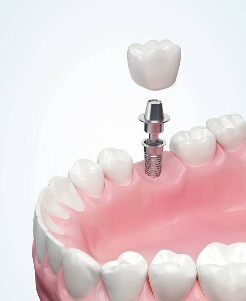
The treated tooth requires a full coverage crown to protect it from fracture. At Cosmolaser, crown options include porcelain-fused-to-metal, full zirconia and E.max ceramic — discussed at consultation or the post-root-canal review.
Treatment | Why It Follows Root Canal | Link |
|---|---|---|
Dental Crowns (Zirconia / E.max) | A crown is the mandatory next step after root canal treatment on posterior teeth — without it, the treated tooth is highly prone to fracture. | /services/dental/veneers-smile-makeover-sharjah/ |
CBCT & OPG Dental Imaging | Our in-house CBCT scanner is used for all root canal cases — no referral needed. Also used for implant planning, oral surgery and orthodontics. | /services/dental/cbct-opg-dental-xray-sharjah/ |
Dental Implants | If a tooth cannot be saved despite root canal treatment — due to vertical fracture or severely compromised structure — implant replacement is planned. | /services/dental/dental-implants-sharjah/ |
Teeth Whitening | Root-canal-treated teeth can discolour over time from internal staining. Internal bleaching and external whitening are available post-treatment. | /services/dental/teeth-whitening-sharjah/ |
Clear Aligners | Following completion of root canal treatment and crown placement, patients often address overall dental alignment as a next aesthetic step. | /services/dental/clear-aligners-sharjah/ |
Veneers & Smile Makeover | Anterior (front) teeth treated with root canal may benefit from veneer placement to restore aesthetics — coordinated with endodontic treatment. | /services/dental/veneers-smile-makeover-sharjah/ |
We’ve gathered the most common questions to help you feel informed, confident, and ready for your journey with us.
With modern local anaesthetics and technique, root canal treatment is not painful for the vast majority of patients. The procedure is performed under local anaesthetic — you will feel pressure and movement but not pain. The discomfort most people associate with root canal treatment is the toothache that brings them to the clinic, not the procedure itself. After treatment, mild soreness for 48–72 hours is normal and easily managed with standard pain relief. Many patients are genuinely surprised at how straightforward the experience is.
This depends on the tooth and clinical situation. Straightforward single-rooted teeth (incisors, canines, premolars) with no active abscess are often completable in a single session of 60–90 minutes. Multi-rooted teeth (molars), teeth with active abscesses, or complex anatomy cases are managed in two sessions spaced 1–2 weeks apart, with antibacterial medication placed between visits. Your dentist will confirm the likely session count at consultation after reviewing your CBCT scan.
A standard 2D dental X-ray only shows the tooth in one plane — it frequently misses additional root canals, curved canal paths, and the full extent of periapical infection. A CBCT 3D scan gives your dentist a complete map of the tooth's root system before any instrument is placed. This means no canals are missed, the treatment plan is accurate, and the procedure is more predictable. At Cosmolaser, CBCT is standard for all root canal cases — no extra referral required.
Yes — for posterior teeth (premolars and molars), a crown is strongly recommended and considered part of the complete treatment. A root-canal-treated tooth becomes more brittle without its blood supply and is at significantly higher risk of fracture under normal biting forces. A crown protects the tooth structure and extends the life of the treatment. For front teeth, a crown may not always be necessary — your dentist will advise based on the amount of remaining tooth structure.
With proper crown placement, good oral hygiene and regular dental check-ups, a root-canal-treated tooth can last a lifetime. Studies consistently show that root-canal-treated teeth with full coverage crowns have long-term survival rates comparable to natural untreated teeth. The most common reasons for failure are crown fracture, new decay around the restoration, or (less commonly) root fracture — all of which are largely preventable with maintenance.
Root canal treatment saves your natural tooth; extraction removes it. Keeping your natural tooth is almost always the preferred clinical outcome — natural teeth function better under biting load, maintain jawbone density, and do not require replacement. Extraction creates a gap that, if not addressed with an implant or bridge, leads to shifting of adjacent teeth, bone loss and bite problems. Root canal treatment, even when followed by crown placement, is typically less expensive than extraction plus implant replacement of the same tooth.
Root canal treatment cost varies by tooth type and complexity. Anterior teeth (incisors, canines) are lower cost due to simpler single-canal anatomy. Posterior teeth (molars) with 3–4 canals or complex anatomy are priced higher. Retreatment cases are priced according to complexity. We provide a clear written cost estimate at consultation — including the cost of the subsequent crown — so you have the full picture before any treatment begins. WhatsApp us to enquire about current pricing.
Avoid eating until the local anaesthetic has fully worn off (typically 2–4 hours) to prevent accidental biting. Once sensation returns, soft foods are recommended for 24–48 hours while any post-procedure tenderness settles. After this period, eat normally — but avoid biting directly on the treated tooth with hard foods until the permanent crown is in place, as the temporary filling is not designed for heavy biting load.
Same-day appointments available for dental emergencies. CBCT-guided root canal treatment at Cosmolaser Medical Centre, Sharjah — pain-free, precise, and designed to save your tooth.