Oral surgeon-led wisdom tooth extraction under local anaesthesia — CBCT nerve mapping, same-day procedure, and expert aftercare at Cosmolaser Medical Centre, Sharjah.
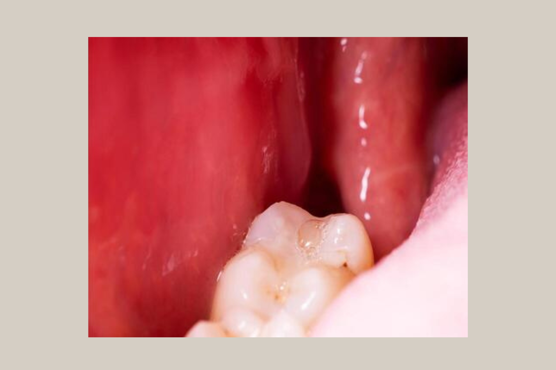
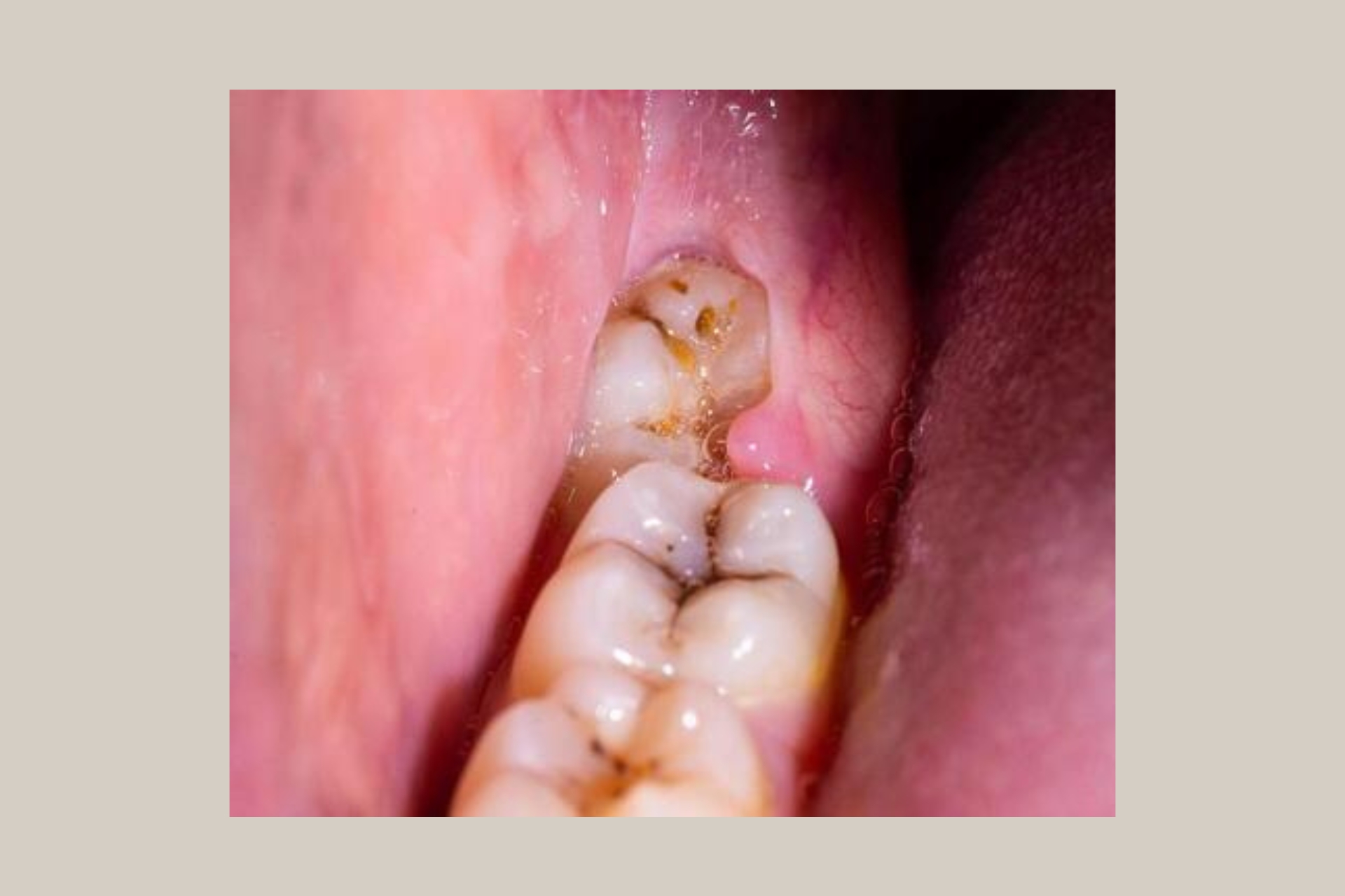
Wisdom teeth — the third molars — typically erupt between the ages of 17 and 25, though in many patients they attempt to erupt well into the 30s. In the UAE’s diverse population, which includes a large proportion of residents in their 20s and 30s from South Asia, the Arab world and Southeast Asia, wisdom tooth problems are among the most common dental presentations seen at Cosmolaser. Jaw size relative to tooth size varies significantly across ethnic groups, and many patients simply do not have adequate arch space for third molars to erupt cleanly.
When a wisdom tooth cannot erupt fully — a condition called impaction — the consequences are predictable and progressive. Partially erupted teeth create a flap of gum tissue (an operculum) that traps food and bacteria, leading to repeated episodes of pericoronitis: painful, swollen infection of the gum around the tooth that can spread to the throat and neck if not treated. Fully impacted teeth exert pressure on the adjacent second molar, causing bone loss, root resorption and decay in the tooth in front. None of these problems resolve without removal.
The key clinical question for lower wisdom teeth is the proximity of the tooth roots to the inferior alveolar nerve (IAN) — the nerve running through the lower jaw that provides sensation to the lower lip and chin. Standard 2D dental X-rays show the tooth and nerve in a single plane, making it impossible to accurately assess the true three-dimensional relationship between root and nerve canal. At Cosmolaser, we use the in-house CBCT scanner to image every lower wisdom tooth in three dimensions before surgery, giving our oral surgeon precise spatial data on nerve proximity, root curvature and bone density. This is not routine practice at most dental clinics in Sharjah — it is the single most important safety measure available for lower wisdom tooth surgery.
Wisdom tooth removal (third molar extraction) is an oral surgical procedure to remove one or more of the four wisdom teeth — the last permanent teeth to develop, located at the very back of the upper and lower arches. The procedure ranges from a straightforward extraction of a fully erupted tooth to a more involved surgical extraction of a deeply impacted tooth requiring bone removal, tooth sectioning or flap reflection.
The procedure is performed under local anaesthesia at Cosmolaser — the surgical site is fully numbed so you feel pressure and movement but no pain. Our oral surgeon determines the surgical approach from the CBCT imaging data: the type of impaction, angulation, root morphology, and proximity to adjacent teeth and anatomical structures (nerve canal, maxillary sinus) are all assessed pre-operatively. A surprise during surgery is a sign of inadequate pre-operative planning — CBCT eliminates most surgical surprises.
Impaction Type | Description | Frequency | Surgical Complexity |
|---|---|---|---|
Mesioangular | Tooth tilted forward towards second molar — most common lower impaction | ~44% of lower impactions | Moderate — often requires sectioning |
Vertical | Upright but insufficient space to erupt fully | ~38% of lower impactions | Low–moderate — may be simple or surgical |
Distoangular | Tooth tilted backward, away from second molar | ~6% of lower impactions | High — angulation complicates delivery |
Horizontal | Tooth lying completely on its side, impacting second molar root | ~3% of lower impactions | High — always requires sectioning and bone removal |
High / Deep impaction | Root apex near or overlapping IAN canal on CBCT | Variable | Very high — nerve proximity requires surgical precision; CBCT mandatory |
Upper wisdom teeth | Usually simpler — no nerve proximity risk; sinus proximity assessed on CBCT | Common | Low–moderate |
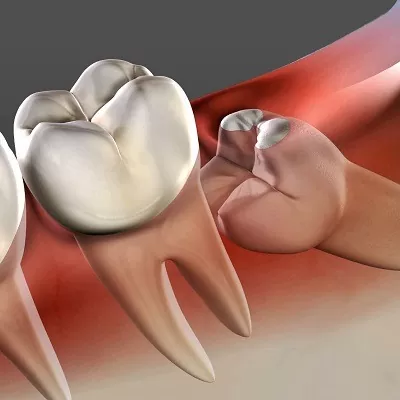
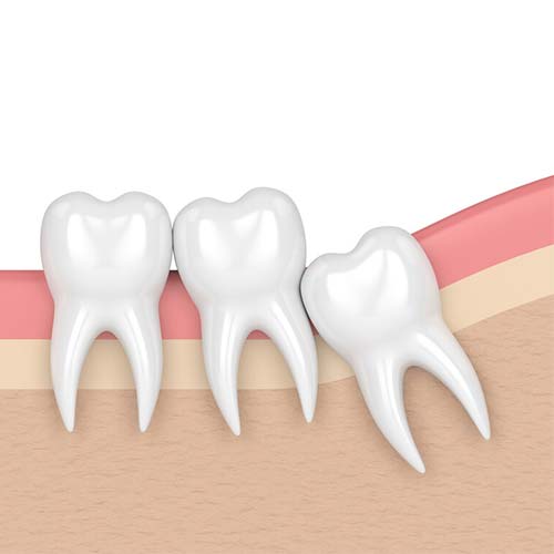
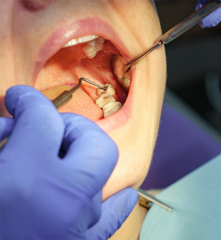
Wisdom tooth extraction sits on a spectrum from routine to genuinely complex oral surgery. A fully erupted, upright wisdom tooth in a patient with adequate space can be removed by an experienced general dental practitioner. A partially or fully impacted lower wisdom tooth with curved roots in close proximity to the inferior alveolar nerve is a surgical procedure that requires specialist oral surgery training, appropriate instrumentation and the judgement that comes from performing hundreds of similar cases.
At Cosmolaser, all wisdom tooth extractions — including straightforward cases — are assessed and performed by our specialist oral surgeon. This means the pre-operative CBCT review, surgical planning, intra-operative decision-making and complication management are in the hands of someone specifically trained for them. Patients referred to us after complications from extractions at other clinics consistently cite the same root cause: inadequate pre-operative imaging and surgical planning.
For wisdom teeth that have fully or sufficiently erupted, with straightforward root anatomy and adequate access, extraction is performed with dental forceps and elevators under local anaesthesia. The tooth is loosened within its socket by expanding the periodontal ligament space, then delivered cleanly. No incision or bone removal is required. Post-operative recovery is typically straightforward — mild soreness for 2–3 days, minimal swelling.
Indication | Fully or mostly erupted wisdom tooth with simple root anatomy |
Anaesthesia | Local anaesthetic — inferior alveolar nerve block (lower) or local infiltration (upper) |
Duration | 15–30 minutes per tooth |
Bone Removal | Not required |
Recovery | Soreness 2–3 days; swelling minimal; return to work next day usually |
CBCT | Required for lower teeth — confirms nerve position and root anatomy before proceeding |
For partially or fully impacted wisdom teeth, surgical extraction is required. The oral surgeon raises a small mucoperiosteal flap (lifts the gum away from the bone), removes a conservative amount of bone overlying the tooth using a surgical handpiece, sections the tooth into two or more pieces if required by the angulation, and delivers each piece separately. The socket is irrigated, the flap repositioned and sutured. Absorbable sutures dissolve within 7–14 days — no suture removal appointment is needed in most cases.
The extent of bone removal and sectioning required is determined entirely by the CBCT data reviewed before the procedure. Our oral surgeon plans the precise surgical approach — flap design, bone removal extent, sectioning pattern — from the 3D image data before entering the surgical field. This pre-operative planning is what distinguishes a controlled, predictable surgical extraction from an improvised one.
Indication | Partially or fully impacted wisdom tooth — any angulation |
Anaesthesia | Local anaesthetic — IAN block with supplemental buccal and lingual infiltrations |
Duration | 30–60 minutes per tooth depending on complexity |
Technique | Flap reflection, bone removal, tooth sectioning (where required), socket irrigation, suturing |
Sutures | Absorbable — dissolve in 7–14 days; no removal appointment needed |
Recovery | Soreness 3–5 days; swelling peaks at 48 hours, resolves by day 5–7; soft diet 5–7 days |
CBCT | Mandatory — full 3D pre-operative planning from imaging data |
Where clinical assessment and patient health allow, all four wisdom teeth can be removed in a single appointment under local anaesthesia. This is the most efficient approach for patients with multiple impacted or symptomatic wisdom teeth, avoiding four separate procedures and recovery periods. The oral surgeon assesses total procedure time, patient tolerance, and the complexity of each individual tooth before confirming a single-session approach is appropriate.
For patients with significant dental anxiety, or where four simultaneous surgical extractions represent a lengthy and physiologically demanding procedure, the teeth may be staged: upper teeth in one session and lower teeth in a separate session one to two weeks later. Both approaches are discussed and the decision is made jointly with the patient.
Session Structure | All four in one visit, or upper/lower staged across two visits |
Duration (all four) | 90–150 minutes depending on individual tooth complexity |
Anaesthesia | Multiple nerve blocks — bilateral IAN blocks (lower) and bilateral infiltrations (upper) |
Recovery | Staged recovery — both sides sore simultaneously; plan 3–5 days off |
Ideal For | Patients wanting to complete treatment in minimum visits; all teeth symptomatic or high-risk |
For lower wisdom teeth where CBCT imaging confirms the root apices are in direct contact with, or wrapping around, the inferior alveolar nerve canal — a situation where full extraction carries a significant risk of permanent nerve damage (paraesthesia or anaesthesia of the lower lip and chin) — coronectomy is an evidence-based alternative. The crown of the tooth is removed down to the cemento-enamel junction, intentionally leaving the roots in situ. The roots, deprived of their blood supply through the crown, undergo predictable bone deposition over them and rarely cause ongoing problems.
Coronectomy is not a compromise — it is the clinically correct treatment for specific nerve-proximity cases, endorsed by NICE guidelines and international oral surgery consensus. It significantly reduces the risk of inferior alveolar nerve injury compared to full extraction in high-risk cases, while eliminating the infectious and pressure risks of the retained crown. The decision to perform coronectomy versus full extraction is made from the CBCT data at the pre-operative planning appointment.
Indication | CBCT-confirmed direct IAN canal contact or root grooving — nerve injury risk unacceptably high |
Technique | Crown section and removal to CEJ level; roots intentionally retained |
Anaesthesia | Local anaesthetic — same as full surgical extraction |
Duration | 30–45 minutes per tooth |
Follow-up | CBCT at 6 and 24 months to confirm root migration away from nerve and bone deposition |
Success Rate | Approx. 95% of retained roots remain stable; ~5% require subsequent removal, usually easier as roots migrate coronally |
CBCT | Mandatory — this decision cannot be made from 2D radiographs |
The oral surgeon’s recommendation follows full CBCT review and clinical assessment. This table maps typical clinical findings to the likely surgical approach:
Your Primary Concern | Recommended Protocol | Expected Outcome |
|---|---|---|
Stubborn belly fat / love handles | Fat Reduction (RF + Ultrasound) | 2–4cm circumference reduction per course |
Loose skin after weight loss or pregnancy | Skin Tightening (RF focused) | Firmer, more elastic skin; improved tone |
Cellulite on thighs or buttocks | Cellulite Protocol (Vacuum + RF) | Smoother skin texture; reduced dimpling |
Overall body reshaping (fat + skin) | Combined Slimming + Contouring | Comprehensive inch loss + tightening |
Post-baby body restoration | Full Protocol (all 3 pillars) | Abdominal tightening, fat reduction, tone |
Arm or thigh definition before an event | Accelerated course (2x/week sessions) | Visible definition in 3–4 weeks |
Raised / hypertrophic scars | Intralesional injection + laser | Different pathway — assess at consultation |
The oral surgeon’s recommendation follows full CBCT review and clinical assessment. This table maps typical clinical findings to the likely surgical approach:
CBCT / Clinical Finding | Surgical Approach | Notes |
|---|---|---|
Fully erupted, simple root form, no impaction | Simple forceps extraction | CBCT still taken for lower teeth to confirm nerve clearance |
Partially erupted, mesioangular impaction | Surgical extraction with sectioning | Most common lower impaction scenario |
Horizontal impaction, abutting second molar | Surgical extraction — bone removal + sectioning | Careful technique to protect second molar root |
Distoangular impaction — high complexity | Surgical extraction with extended access | Most surgically demanding angulation; planned in detail from CBCT |
Deep vertical impaction, straightforward roots | Surgical extraction with bone removal | Depth requires more bone access; roots away from nerve |
Root apices overlapping IAN canal on CBCT | Coronectomy | Full extraction carries unacceptable nerve injury risk; coronectomy is correct treatment |
Upper wisdom tooth — standard impaction | Surgical extraction (no nerve risk) | Sinus proximity assessed on CBCT; Schneiderian membrane protection where needed |
Acute pericoronitis (active infection) | Antibiotics first, then extraction in 5–7 days | Inflamed tissue reduces anaesthetic efficacy; operating through active infection increases risk |
Patient with high dental anxiety | All approaches available — discuss sedation | Oral sedation option discussed at consultation |
The inferior alveolar nerve (IAN) runs through a canal inside the lower jaw, and the roots of lower wisdom teeth develop in close proximity to — or in some cases directly adjacent to, or grooved around — this canal. Damage to the IAN during extraction can cause temporary or permanent numbness, tingling or altered sensation of the lower lip, chin and teeth on the affected side. Temporary paraesthesia resolves in most cases within weeks to months; permanent paraesthesia is a life-altering complication that cannot be reversed.
A standard 2D periapical X-ray shows the tooth and nerve canal as overlapping shadows in a single plane. When the nerve canal appears to cross the root on a 2D film, it may be passing in front of or behind the root (safe), or directly through the root itself (high risk) — a 2D image cannot tell you which. CBCT resolves this completely: the three-dimensional relationship between root and nerve canal is unambiguous in axial, coronal and sagittal slices.
Standard 2D Periapical X-ray | In-house CBCT 3D Scan (Cosmolaser) |
|---|---|
Cannot determine true root-to-nerve spatial relationship | Exact 3D relationship between root apices and IAN canal mapped |
Overlapping shadows — nerve position ambiguous | Axial, coronal and sagittal views resolve all ambiguity |
Root curvature often underestimated | Full root form and curvature visible in all planes |
Cortication of nerve canal assessed only in one plane | Loss of cortication (root invasion) detectable pre-operatively |
Cannot distinguish nerve passing in front vs through root | Buccal, lingual or direct nerve contact identified precisely |
Coronectomy decision cannot be safely made | Coronectomy planned with confidence from CBCT data |
Available at most dental clinics | In-house at Cosmolaser — same visit, instant review |
At Cosmolaser, CBCT imaging is performed for every lower wisdom tooth extraction before a surgical plan is confirmed. This is not routine practice at most dental clinics in Sharjah — it is the standard we hold ourselves to because the consequences of not imaging in 3D are borne by the patient, not the surgeon.
Male patient, 24 years. Bilateral lower wisdom tooth pain and repeated pericoronitis episodes. Both lower wisdom teeth mesioangularly impacted on OPG.
| Findings | ||
|---|---|---|
|
CBCT bilateral: both root apices within 2–3mm of IAN canal — not overlapping. Mesioangular impaction both sides. Roots straight, single curvature. No cortication loss of IAN canal. |
Treatment: Both lower wisdom teeth removed in same session under bilateral IAN blocks. Flap reflection, buccal bone removal, mesial sectioning, controlled delivery. Absorbable sutures. Prescribed ibuprofen + amoxicillin. |
Outcome: Uneventful recovery. Full sensation confirmed at 1-week review. No pericoronitis recurrence. OPG confirms complete root removal. |
Female patient, 31 years. Right lower wisdom tooth pain. Partially impacted. OPG showed darkening of roots at IAN canal level — high-risk appearance.
| Findings | ||
|---|---|---|
|
CBCT: right lower wisdom tooth roots grooved around IAN canal with loss of cortication on lingual aspect. Full extraction assessed as carrying high risk of permanent paraesthesia.
|
Treatment: Coronectomy performed. Crown sectioned and removed to CEJ level. Roots intentionally retained. Socket irrigated. Primary closure. CBCT follow-up scheduled at 6 and 24 months. |
Outcome: No nerve symptoms post-operatively. 6-month CBCT: retained roots migrated 1.5mm coronally away from nerve canal. Bone deposition confirmed. No symptoms. |
Male patient, 27 years. Dull lower left jaw ache. No pericoronitis history. OPG showed horizontal lower left wisdom tooth impacting second molar root.
| Findings | ||
|---|---|---|
|
CBCT: horizontal impaction confirmed. Root apex 4mm from IAN canal. Second molar root shows early external resorption at point of contact. No active infection. |
Treatment: Urgent surgical extraction recommended to prevent further second molar root damage. Extended flap, significant buccal bone removal, horizontal sectioning into 3 pieces. Delivered piece by piece. Sutures placed. |
Outcome: Second molar root resorption arrested. No nerve symptoms. Extraction confirmed complete on OPG. Second molar vitality preserved — no root canal required. |
Female patient, 22 years. All four wisdom teeth partially erupted, recurrent pericoronitis upper right and lower left. Patient requested single-session removal.
| Findings | ||
|---|---|---|
|
CBCT both lower teeth: adequate nerve clearance confirmed. Upper teeth: sinus proximity assessed — both upper roots clear of maxillary sinus floor. Complexity assessed as moderate for all four. |
Treatment: All four removed in single 110-minute session. Upper teeth simple surgical extractions. Lower teeth surgical with sectioning. Bilateral IAN blocks and bilateral upper infiltrations. Total 8 local anaesthetic cartridges. |
Outcome: Patient managed well throughout. No intra-operative complications. Recovery as expected — bilateral swelling day 2–3, resolved by day 6. Full sensation both sides confirmed at 1 week. |
Female patient, 35 years. Right upper wisdom tooth pain. GP dentist declined to extract, citing ‘sinus risk’, and referred to specialist.
| Findings | ||
|---|---|---|
|
CBCT: upper right wisdom tooth root within 1mm of maxillary sinus floor — direct sinus proximity. No root penetration of sinus. Upright impaction, adequate access. |
Treatment: Surgical extraction with sinus-aware technique. Slow, controlled delivery. Immediate post-extraction socket inspection confirmed sinus membrane intact. No oro-antral communication. Socket dressed with collagen. |
Outcome: No sinus complication. No oro-antral communication. 1-week review: normal healing. Patient satisfied — avoided hospital referral. |
Male patient, 19 years. Severe right lower jaw pain, facial swelling, trismus — mouth opening limited to 18mm. Temperature 38.1C. Emergency presentation.
| Findings | ||
|---|---|---|
|
CBCT: partially impacted lower right wisdom tooth with large pericoronitis cavity. No deep space infection spread — confined to pericoronary region. IAN canal clear. |
Treatment: Antibiotics prescribed (amoxicillin + metronidazole) and systemic analgesia. Extraction deferred 5 days until infection resolved. Wisdom tooth removed at day 6 under IAN block once opening restored to 35mm. |
Outcome: Full infection resolution before surgery. Uneventful extraction at follow-up. No recurrence. Patient counselled on opposite side lower wisdom tooth monitoring. |
Clinical examination including mouth opening assessment, palpation of the affected area, and inspection of gum health around the wisdom tooth. OPG panoramic X-ray taken for overall assessment. CBCT scan of the relevant quadrant(s) for all lower wisdom teeth and upper teeth near the sinus. Oral surgeon reviews CBCT chairside and explains findings, impaction type, nerve relationship and recommended approach. Written cost estimate provided.
Eat a normal light meal 2–3 hours before your appointment — do not attend on an empty stomach. Take any regular medications as normal unless specifically advised otherwise. Arrange a driver if you feel you may need assistance — most patients drive themselves home, but surgical procedures can leave some patients feeling light-headed. Wear comfortable clothing. Avoid alcohol for 24 hours before surgery.
Topical anaesthetic gel applied to gum before injection. Local anaesthetic administered — inferior alveolar nerve block for lower teeth (numbs the entire lower half of the face on that side), local infiltration for upper teeth. The oral surgeon confirms complete anaesthesia before proceeding — you feel pressure and movement throughout but should feel no pain. If you do, raise your hand immediately and additional anaesthetic will be given.
Surgical procedure performed as planned from CBCT data. Simple extractions: forceps and elevator delivery. Surgical extractions: flap, bone removal, sectioning, delivery, socket irrigation. The oral surgeon explains what you will feel at each stage. You are awake and conscious throughout — local anaesthesia is highly effective and general anaesthesia is not required for the vast majority of wisdom tooth extractions.
Socket inspected for complete root and bone removal. Irrigated with sterile saline. Haemostatic dressing placed if needed. Absorbable sutures placed for surgical extractions — they dissolve in 7–14 days. Gauze pack placed over extraction site and patient asked to bite firmly for 30–40 minutes to aid clot formation.
Bite on gauze for 40 minutes. No rinsing, spitting or smoking for 24 hours — this dislodges the clot and causes dry socket. Cold compress (ice pack wrapped in cloth) applied to face externally for 20 minutes on/off for the first 6 hours. Soft diet for 5–7 days. Ibuprofen and paracetamol alternated for pain management — prescription pain relief provided if needed. Antibiotics prescribed where infection risk is elevated. Oral hygiene maintained gently — gentle warm saline rinses from day 2.
Sutures inspected (dissolving, no removal needed). Sensation confirmed — lower lip and chin. Oral hygiene guidance for the healing socket. Any complications (dry socket, infection, persistent swelling) assessed and managed.
Dry socket (alveolar osteitis) occurs in approximately 5% of wisdom tooth extractions — more commonly in lower teeth, smokers and patients who did not follow post-operative instructions. Symptoms: worsening pain from day 3–4, empty socket, foul odour. Treatment: socket irrigation and medicated dressing placed by the surgeon, replaced every few days until healing progresses. Pain resolves rapidly after dressing.
Treatment | How It Connects | Link |
|---|---|---|
CBCT & OPG Imaging | Mandatory for all lower wisdom teeth at Cosmolaser — in-house, same-day, instant review. Also | /services/dental/cbct-opg-dental-xray-sharjah/ |
images sinus proximity for uppers. | ||
Dental Implants | Where wisdom tooth extraction creates a gap requiring restoration, or where an adjacent second molar has been compromised, implant planning is integrated. | /services/dental/dental-implants-sharjah/ |
Root Canal Treatment | Adjacent second molars damaged by impacted wisdom teeth may require root canal treatment — assessed and managed in-house. | /services/dental/root-canal-sharjah/ |
Dental Braces | Post-wisdom tooth removal is often the right time to address crowding that wisdom teeth were worsening — orthodontic assessment recommended. | /services/dental/braces-sharjah/ |
Clear Aligners | Aligner treatment is often started after wisdom teeth are removed — removes the risk of new crowding during treatment. | /services/dental/clear-aligners-sharjah/ |
TMJ Treatment | Difficult or prolonged wisdom tooth procedures can temporarily stress the TMJ — assessment available if jaw pain develops post-extraction. | /services/dental/tmj-sharjah/ |
We’ve gathered the most common questions to help you feel informed, confident, and ready for your journey with us.
The procedure itself is performed under local anaesthesia and should not be painful — you will feel pressure, pushing and movement, but not pain. The inferior alveolar nerve block used for lower wisdom teeth is highly effective and numbs the entire lower half of the face on that side. If at any point during the procedure you feel pain rather than pressure, raise your hand and additional anaesthetic will be administered immediately. Post-operative soreness begins as the anaesthetic wears off (2–4 hours after the procedure) and is well managed with alternating ibuprofen and paracetamol.
No — the vast majority of wisdom tooth extractions, including complex surgical extractions of fully impacted lower wisdom teeth, are safely and comfortably performed under local anaesthesia in a dental chair. General anaesthesia is generally only required for patients with severe dental phobia where conscious treatment is not feasible, medical conditions requiring monitored anaesthesia, or very young children. GA for routine wisdom tooth removal is not necessary and carries its own risks. Our oral surgeon's skill with local anaesthetic technique means most patients tolerate even difficult surgical extractions with minimal discomfort.
A 2D X-ray cannot tell your surgeon whether the inferior alveolar nerve — the nerve controlling sensation in your lower lip and chin — is passing in front of, behind, or directly through your wisdom tooth roots. This is critical safety information: if the nerve is in direct contact with the roots and the surgeon does not know, the risk of permanent nerve damage is significantly elevated. A CBCT 3D scan resolves this completely, showing the exact spatial relationship between root and nerve in every plane. At Cosmolaser this is performed in-house at your consultation appointment and reviewed immediately.
A coronectomy removes only the crown of the wisdom tooth, intentionally leaving the roots in place when their removal would carry an unacceptably high risk of damaging the inferior alveolar nerve. The retained roots are gradually covered by bone and typically cause no ongoing problems. This is not a compromise — it is the clinically correct, evidence-based treatment for specific high-risk nerve proximity cases, endorsed by international oral surgery guidelines. The decision is made from CBCT data at the pre-operative planning appointment. Cosmolaser is one of very few clinics in Sharjah offering coronectomy as a planned procedure rather than referring these patients to hospital.
For simple extractions: mild soreness for 2–3 days, minimal swelling, return to normal activities the following day. For surgical extractions of impacted teeth: soreness 3–5 days, swelling peaks at 48 hours and resolves by day 5–7, soft diet for 5–7 days, most patients take 2–3 days off work. For all four teeth removed simultaneously: expect bilateral swelling and a full week of recovery. The socket takes 6–8 weeks to fully fill with bone, though the surface heals within 2–3 weeks. Sensible soft diet, good oral hygiene and no smoking significantly reduce recovery time.
Dry socket (alveolar osteitis) occurs when the blood clot in the extraction socket is dislodged or dissolves prematurely, exposing the bare bone and causing severe, throbbing pain from day 3–4 onwards. It affects approximately 5% of extractions, more commonly lower teeth and patients who smoke. To avoid it: do not rinse, spit forcefully or smoke for 24 hours after extraction. Avoid straws. Keep pressure on the gauze pack for the full 40 minutes. If you develop increasing pain from day 3 onwards rather than improving pain, contact us — dry socket is easily treated with a medicated socket dressing that provides rapid relief.
Yes — where the complexity of each tooth allows it and patient health permits, all four wisdom teeth can be removed in a single session. This eliminates the need for four separate procedures and recovery periods. The total procedure time is typically 90–150 minutes. You will have bilateral swelling and soreness post-operatively, so plan for 3–5 days of recovery. The oral surgeon assesses suitability for single-session removal at your consultation after reviewing your CBCT imaging.
Cost varies by the number of teeth, complexity of impaction and surgical approach required. Simple erupted tooth extraction is the lowest cost; full surgical extraction of a deeply impacted lower tooth with bone removal and sectioning is higher. Coronectomy is priced as a surgical procedure. The consultation fee includes your CBCT scan and written cost estimate — so you have the full picture before committing to treatment. WhatsApp us for current pricing and to discuss your specific situation.
Oral surgeon-led wisdom tooth removal with in-house CBCT nerve mapping at Cosmolaser Medical Centre, Sharjah. Safe, precise, same-day where possible.