
The standard process at most dental clinics in Sharjah for complex cases: the dentist examines you, determines a scan is needed, writes a referral to an external imaging centre, you book a separate appointment, attend on a different day, collect the scans, bring them back to the clinic, then return for your treatment planning appointment. That process takes days or weeks — and introduces communication gaps between the imaging centre and your treating dentist.
At Cosmolaser, the CBCT scanner is in the same building as your dentist. At your initial consultation, if a scan is indicated, it is taken immediately — in the same appointment. Your dentist reviews the 3D images while you are still in the chair, discusses the findings with you directly, and produces a complete treatment plan in a single visit. For patients considering dental implants, clear aligners, or complex root canal treatment, this means a definitive plan — with realistic timelines, costs, and clinical detail — on the same day you walk in.
Most Sharjah Dental Clinics | Cosmolaser — In-House CBCT |
|---|---|
Day 1: Consultation — referral written | Day 1: Consultation + scan in same visit |
Day 3+: Attend external imaging centre | Day 1: 3D images reviewed with dentist |
Day 5+: Collect scan, return to clinic | Day 1: Treatment date scheduled |
Day 7+: Treatment planning appointment | Day 1: Treatment date scheduled |
7+ days before treatment can begin | Complete plan in a single visit |
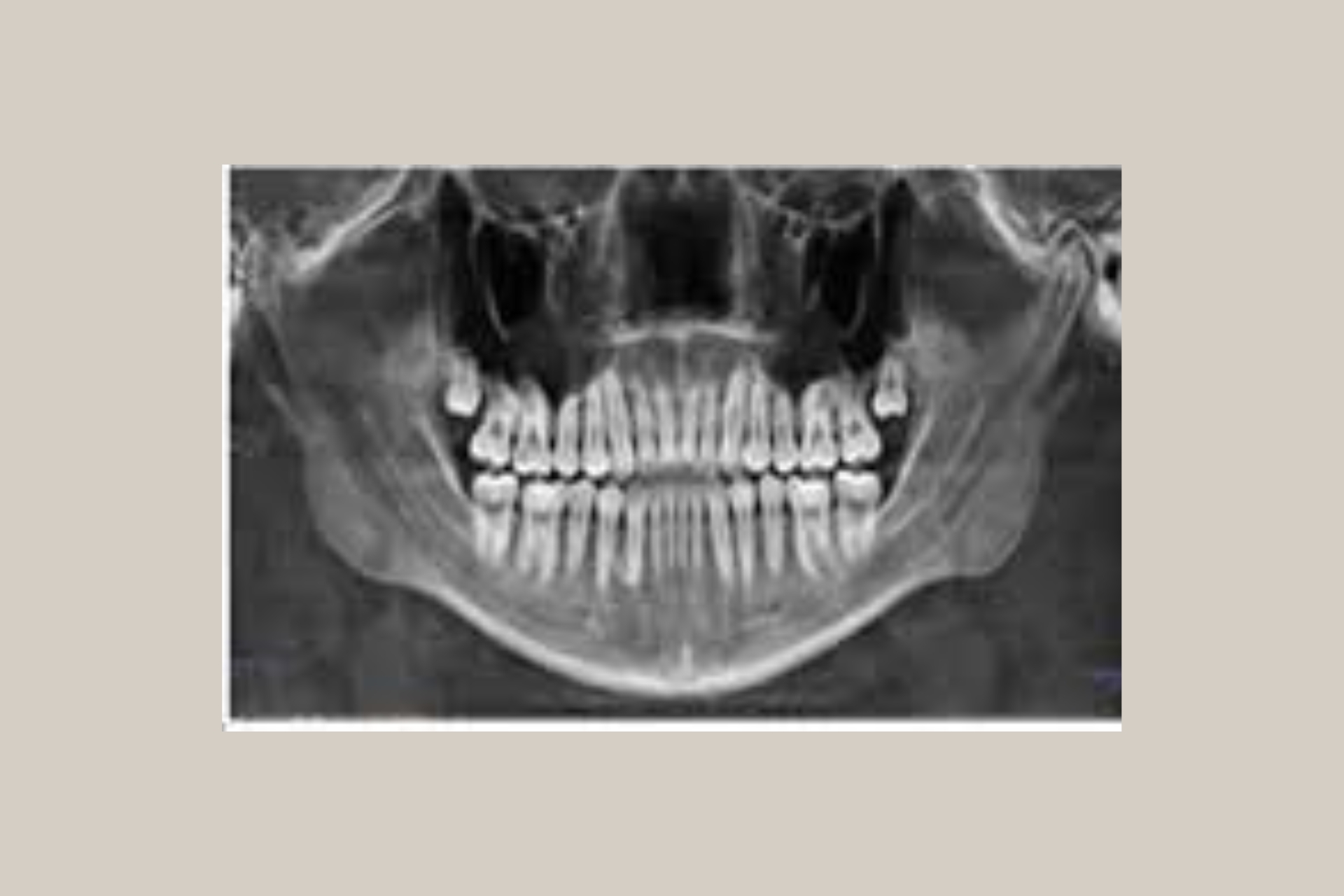
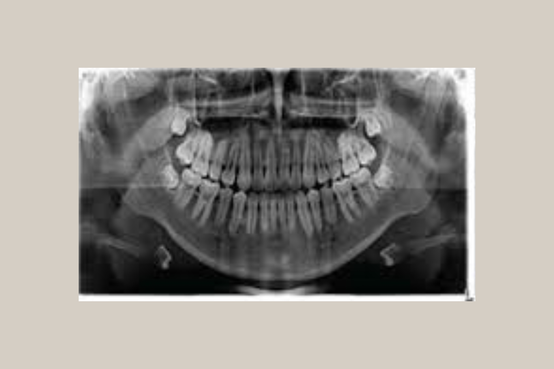
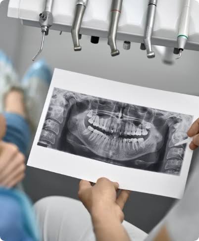
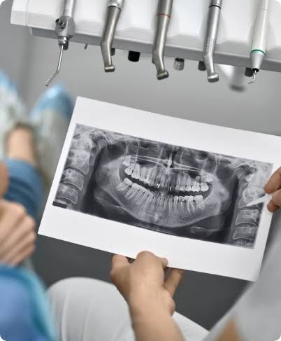
An OPG (Orthopantomogram) is a panoramic dental X-ray that captures the entire upper and lower jaw, all teeth, both temporomandibular joints (TMJ), and the surrounding bone structures in a single 2D image. It is the standard first-step imaging study for new dental patients and is required before most planned dental procedures.
Unlike individual periapical X-rays that focus on one or two teeth at a time, an OPG provides a comprehensive overview of the entire oral and maxillofacial region in one scan — making it the most time-efficient and clinically comprehensive 2D dental imaging study available.
OPG Details | ||
|---|---|---|
Scan type | 2D panoramic X-ray — full arch, both jaws, TMJ joints | |
Duration | Under 60 seconds of actual scan time. Total appointment: 10–15 minutes including preparation. | |
Radiation dose | Very low — approximately equivalent to a few hours of natural background radiation. Significantly lower than a medical CT scan. | |
Preparation | No preparation required. Remove metal jewellery, hair accessories, and glasses before the scan. | |
Result | Digital image available immediately — reviewed with your dentist at the same appointment. | |
Indications | New patient comprehensive assessment · Implant preliminary planning · Orthodontic assessment · Wisdom tooth evaluation · TMJ assessment · Routine monitoring · Pre-surgical planning |
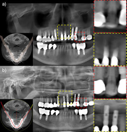
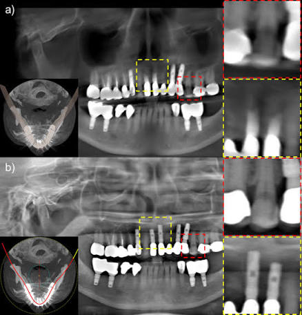
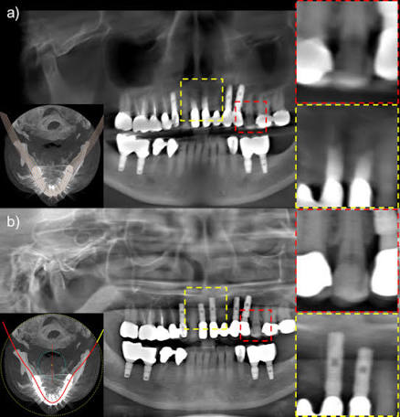
A CBCT (Cone Beam Computed Tomography) scan produces a full three-dimensional model of the teeth, jawbone, nerve canals, sinuses, and surrounding structures — from every angle and at every depth. Where an OPG provides a flat, two-dimensional overview, a CBCT provides a volumetric dataset that can be sliced, rotated, and measured in any plane the clinician requires.
For procedures where the precise three-dimensional position of a tooth, a nerve, or a piece of bone determines the difference between success and failure — dental implants, complex root canal treatment, impacted wisdom tooth removal, orthodontic planning for impacted canines, and orthognathic surgery — a CBCT is not an optional upgrade. It is the standard of care.
Clinical Need | OPG (2D) | CBCT (3D) |
|---|---|---|
Bone volume for implant placement | Approximate only — no depth measurement | ✓ Exact height, width and density in 3 planes |
Nerve canal position | Approximate 2D position | ✓ Exact 3D position — critical for implant safety |
Sinus floor height | Approximate | ✓ Exact — essential for upper molar implants |
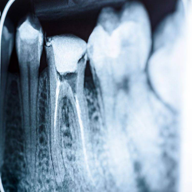
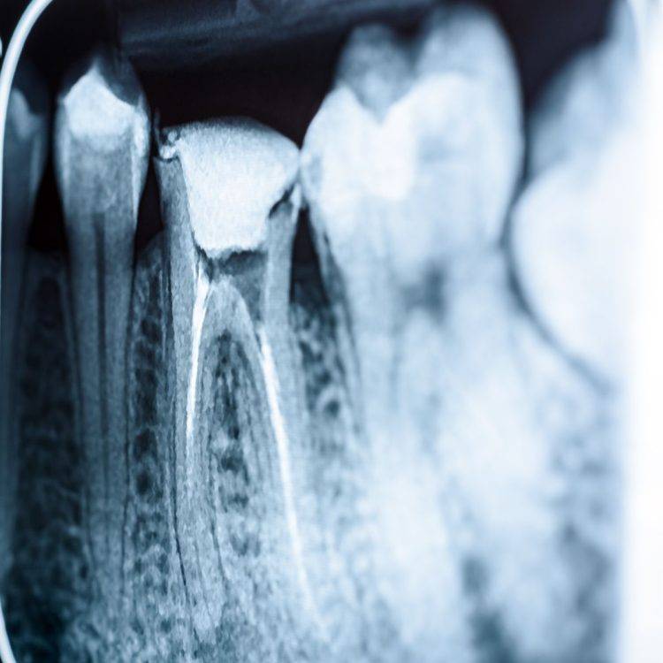
Root canal anatomy | Single 2D projection | ✓ Full 3D — reveals additional canals invisible on OPG |
Impacted tooth depth and angulation | 2D overview | ✓ Exact 3D position relative to adjacent structures |
Jaw cyst or tumour extent | Outlines visible lesion | ✓ Full 3D extent — surgical planning accuracy |
TMJ joint morphology | Limited detail | ✓ Detailed 3D joint surface and condyle anatomy |
Airway assessment | Not possible | ✓ Airway volume and obstruction — for sleep apnoea planning |
CBCT Details | ||
|---|---|---|
Scan type | 3D cone beam CT — full volumetric dataset in multiple planes | |
Duration | 20–40 seconds of scan rotation. Total appointment: 15–20 minutes including preparation and positioning. | |
Radiation dose | Higher than OPG but significantly lower than a conventional medical CT scan. Field of View (FOV) is selected to the minimum area needed for the clinical indication — reducing dose further. | |
Preparation | No preparation required. Remove metal jewellery, hair accessories, and glasses. Inform the radiographer if you are or may be pregnant. | |
Result | 3D images processed and available for review within minutes — at the same appointment at Cosmolaser. | |
Field of View | Small FOV (single tooth/quadrant) · Medium FOV (single arch) · Large FOV (full craniofacial) — selected based on clinical indication to minimise radiation. | |
Indications | Dental implant planning · Complex root canal (endodontics) · Impacted wisdom tooth or canine · Orthodontic treatment planning · Jaw cyst/lesion assessment · TMJ evaluation · Orthognathic surgery planning · Sleep apnoea assessment |
Protocol | Treatments Combined
| Why |
|---|---|---|
New patient with no recent dental records | OPG | Comprehensive baseline — reveals all issues, including asymptomatic ones |
Dental implant planning — any site | CBCT | Bone volume, density, nerve position, and sinus anatomy in 3D |
Upper molar implants (near sinuses) | CBCT | Precise sinus floor height — determines if sinus lift is needed |
Wisdom tooth extraction — lower jaw | OPG + Ceph | OPG for overview; CBCT if nerve proximity is suspected |
Orthodontic treatment — braces or aligners | OPG + Ceph | Tooth positions + skeletal relationship essential for treatment planning |
Impacted canine or premolar | CBCT | Exact 3D position relative to roots and bone — surgical planning |
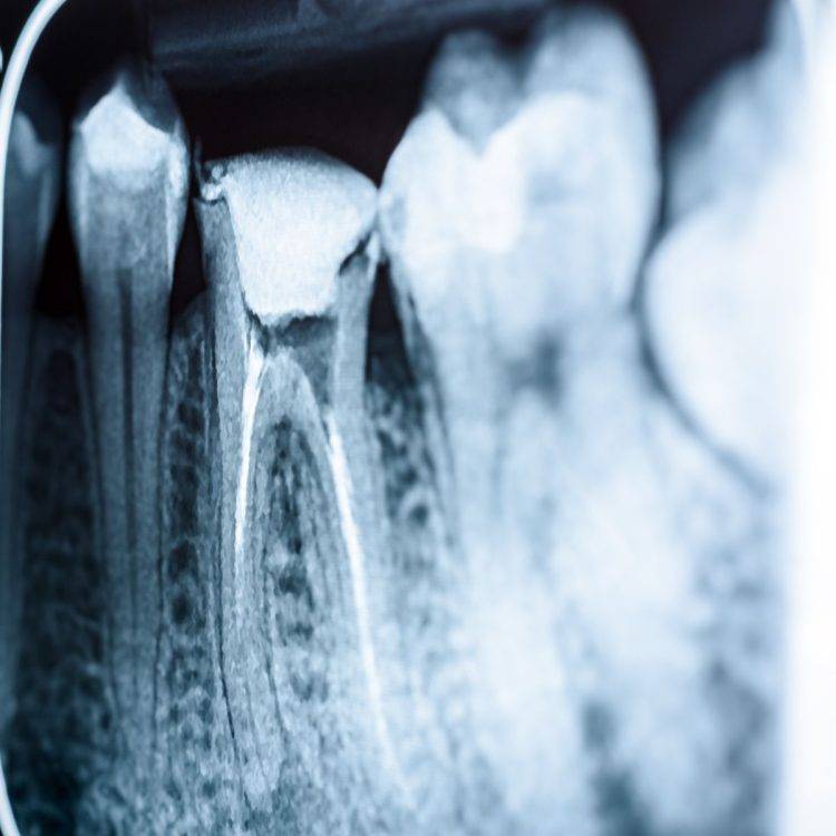
Root canal treatment — multi-rooted teeth | CBCT | Reveals extra canals and root curvatures invisible on periapical X-ray |
Failed previous root canal — retreatment | CBCT | Identifies missed canals, fractures, or perforations |
Jaw pain, clicking, or restricted opening (TMJ) | CBCT | 3D condyle morphology — joint assessment cannot be done adequately on OPG |
Bone grafting — assessment and planning | CBCT | Defect volume and recipient site anatomy in 3D |
Jaw cyst, lesion, or suspected pathology | CBCT | 3D extent of lesion — surgical access and safety planning |
Orthognathic (jaw) surgery planning | CBCT | Full 3D craniofacial data — essential for surgical simulation |
Trauma — suspected jaw fracture | CBCT | Fracture line identification and displacement assessment |
You will be asked to remove any metal items — earrings, necklaces, hair clips, and glasses — as these can create artefacts on the scan. A lead apron will be provided. If you are or may be pregnant, inform the radiographer before the scan is taken.
You will stand (or sit) with your chin resting on a support. Your teeth will gently bite a small, disposable plastic guide that keeps your jaw in the correct position. The technician will position the machine carefully. You hold still — the machine does the moving.
The OPG machine rotates slowly around your head, capturing the panoramic image. You will hear a gentle hum. The exposure itself takes under 20 seconds. You will not feel anything.
For a CBCT, you will be seated or standing inside the CBCT unit. Your head is positioned and stabilised using bite guides and head rests. Correct positioning is important for image quality — your radiographer will take a moment to ensure precise alignment.
The cone beam rotates around your head, capturing data from multiple angles. The scan itself takes under a minute. You will not feel anything.
CBCT images are processed by the software and available for review within minutes. OPG images are available immediately.
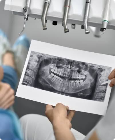
At Cosmolaser, your dentist reviews the images directly with you. You will see your own scan on-screen, and your dentist will explain what it shows — in plain language, not clinical jargon. Your treatment plan is then discussed, with exact clinical detail only possible because of the 3D data.
Imaging Type | Approximate Dose | Equivalent To |
|---|---|---|
Single periapical X-ray | ~0.005 mSv | Less than 1 hour of natural background radiation |
OPG panoramic X-ray | ~0.01–0.02 mSv | A few hours of natural background radiation |
CBCT — small FOV | ~0.05–0.1 mSv | 1–2 days of natural background radiation |
CBCT — medium FOV | ~0.1–0.2 mSv | A few days of natural background radiation |
Medical CT scan (head) | ~2 mSv | ~100× more than a dental CBCT |
Transatlantic flight (return) | ~0.1 mSv | Comparable to a medium FOV dental CBCT |
Annual background radiation (UAE) | ~2–3 mSv | — |
We’ve gathered the most common questions to help you feel informed, confident, and ready for your journey with us.